-
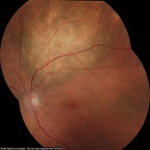
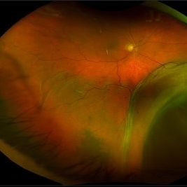
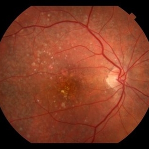
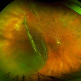
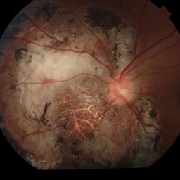
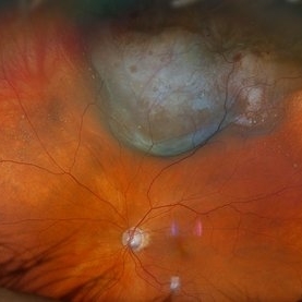
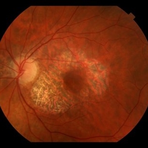
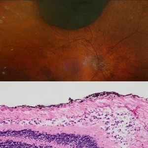
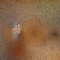
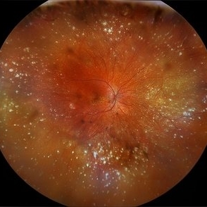
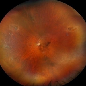
 ERM with Retinal Striae
Apr 13 2023 by Virginia Gebhart
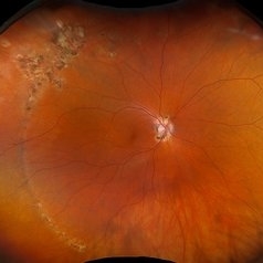
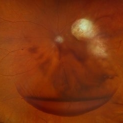
Right eye fundus photo of 65-year-old male with severe ERM with retinal striae s/p bilateral RD repair
Photographer: Virginia Gebhart, Retina Consultants of Carolina
Imaging device: Topcon TRC 50DX
Condition/keywords: epiretinal membrane (ERM), retinal strial
-
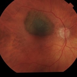
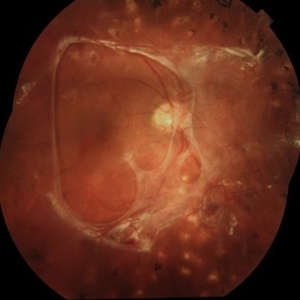
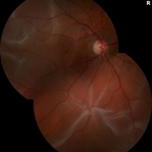
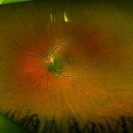
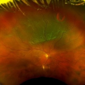
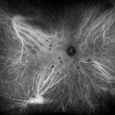
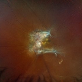
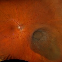
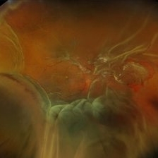
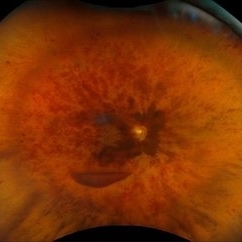
 Central Retinal Vein Occlusion
Apr 13 2023 by Virginia Gebhart
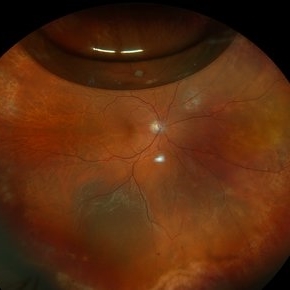
68-year-old male with Central Retinal Vein Occlusion with Macular Edema. Pt presented with VA of count fingers @ 5 ft. Pt was treated with Avastin
Photographer: Virginia Gebhart, Retina Consultants of Carolina
Imaging device: Topcon TRC 50DX
Condition/keywords: central retinal vein occlusion (CRVO)
-
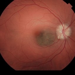
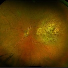
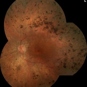
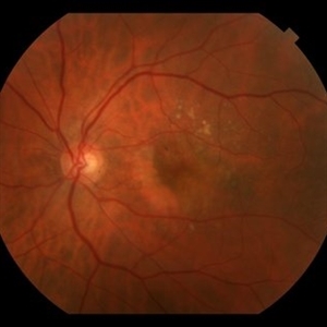
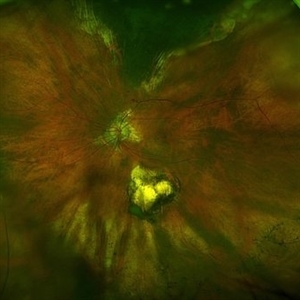
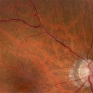
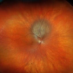
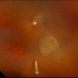
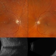
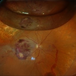
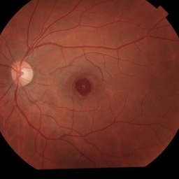
 Stargardt's Disease
May 5 2023 by Virginia Gebhart
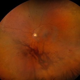
51-year-old male with bilateral central retinal dystrophy consistent with Stargardt disease. No significant progression of central atrophy, and VA has remained stable at 20/150 since 2012
Photographer: Virginia Gebhart, Retina Consultants of Carolina
Imaging device: Topcon TRC 50DX
Condition/keywords: Stargardt disease
-
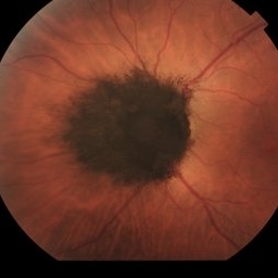
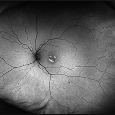
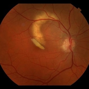
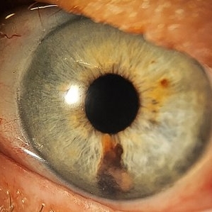
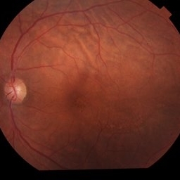
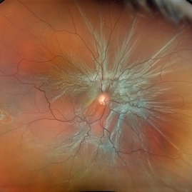
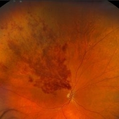
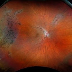
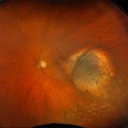
 Syphilitic Uveitis
May 26 2023 by Virginia Gebhart
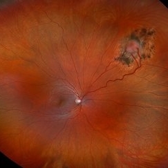
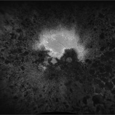
35-year-old female with resolved syphilitic uveitis OU. Severe retinal thinning with chalky ON pallor and severe vessel attenuation associated with presumed post syphilitic neuritis vs bilateral CRAO's in both eyes. Pt reports acute bilateral vision loss after delivery of her child in 8/2021. Limited VA OU due to severe optic nerve and macula atrophy.
Photographer: Virginia Gebhart, Retina Consultants of Carolina
Imaging device: Topcon TRC 50DX
Condition/keywords: syphilis, uveitis
-
 MIDD
MIDD
May 26 2023 by Virginia Gebhart
51-year-old female with dry AMD, advanced atrophic without subfoveal involvement OU. Genetic testing confirmed MIDD (maternal inherited diabetes and deafness) which is a mitochondrial inherited dystrophy. Unaware of any family hx of macular degeneration.
Photographer: Virginia Gebhart, Retina Consultants of Carolina
Imaging device: Topcon TRC 50DX
Condition/keywords: advanced geographic atrophy, geographic atrophy
-
 Stargardt Disease
May 30 2023 by Virginia Gebhart
Pattern dystrophy OU, possible Stargardt disease. Genetic testing done in office to confirm. Vision limited to 20/200 OU due to atrophy
Photographer: Virginia Gebhart, Retina Consultants of Carolina
Imaging device: Topcon TRC 50DX
Condition/keywords: pattern dystrophy, Stargardt Disease
-
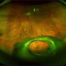
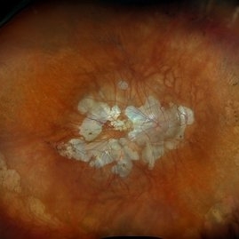
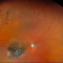
 Regressing Choroidal Melanoma with Asteroid Hyalosis
Jun 2 2023 by Virginia Gebhart
73-year-old female with Asteroid Hyalosis and a stable regressing tumor OD, five months s/p brachytherapy. There is still overlying fluid on exam which should continue to resolve. Vision is stable.
Photographer: Virginia Gebhart, Retina Consultants of Carolina
Imaging device: Optos California
Condition/keywords: asteroid hyalosis
-
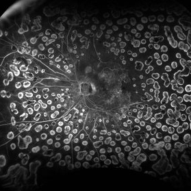
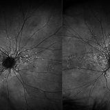
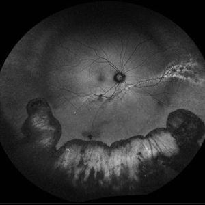
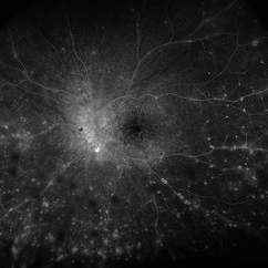
 Retinitis Pigmentosa
Oct 12 2023 by Virginia Gebhart
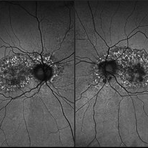
Fundus Auto-Fluorescence photo of 73-year-old woman with Retinitis Pigmentosa, first diagnosed 23 years ago. Extensive outer retinal atrophy with minimal foveal sparing, bone spicule pigmentation and waxy pallor. Vision NLP
Photographer: Virginia Gebhart, Retina Consultants of Carolina
Imaging device: Optos
Condition/keywords: retinitis pigmentosa, retinitis pigmentosa (RP) dystrophy
-
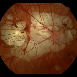
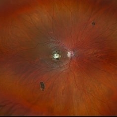
 Dislocated IOL
Oct 12 2023 by Virginia Gebhart
Fundus photo of an 83-year-old man with a 3 piece dislocated IOL. Surgery performed, PPV/removal of nonmagnetic FB/secondary Akreos. Eye is stable, vision limited due to grade 3 VH
Photographer: Virginia Gebhart, Retina Consultants of Carolina
Imaging device: Optos
Condition/keywords: dislocated intraocular lens (IOL), dislocated lens
-
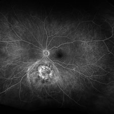
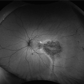
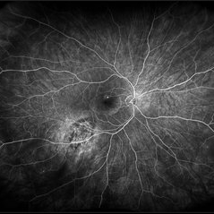
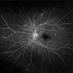
 Severe NPDR
Oct 24 2023 by Virginia Gebhart
Fluorescein angiogram of left eye in 60-year-old male with severe non-proliferative diabetic retinopathy with extensive macular edema. Most recent A1c is 11. Vision 20/400. Injection of Eylea given
Photographer: Virginia Gebhart
Imaging device: Topcon
Condition/keywords: diabetic macular edema, Diabetic Retinopathy, fluorescein angiogram (FA), Fluorescein angiography
-
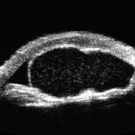 Idiopathic Iris Cyst
Oct 25 2023 by Virginia Gebhart
UBM of recurring idiopathic iris cyst in 72 year old female
Photographer: Virginia Gebhart
Imaging device: Ellex Eye Cubed
Condition/keywords: anterior chamber, cyst, immersion ultrasound, iris
-
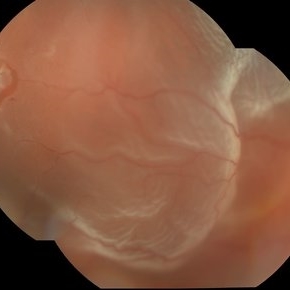
 Retinal Detachment
Oct 26 2023 by Virginia Gebhart
74 year old male with mac-off retinal detachment with single break
Photographer: Virginia Gebhart
Imaging device: Optos
Condition/keywords: detachment, Retinal Detachment, retinal detachment of the macula
-
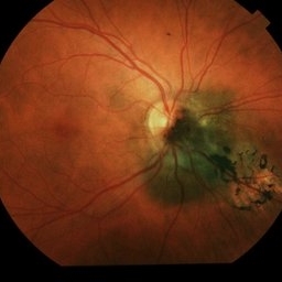
 Choroidal Melanoma
Oct 27 2023 by Virginia Gebhart
76 year old male with suspicious pigmented choroidal lesion with new collar button growth. Blocking defect and vascularity noted on FA
Photographer: Virginia Gebhart
Condition/keywords: FA late phase, fluorescein angiogram (FA), Fluorescein angiography, melanoma
-
 Gore Tex Suture
Nov 1 2023 by Virginia Gebhart
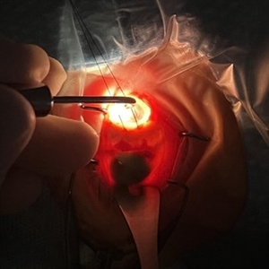
Gore Tex suture of scleral fixated lens in 66 year-old male, 1 day post-op
Photographer: Virginia Gebhart
Imaging device: slit lamp camera
Condition/keywords: Gore Tex Suture, post-op, Scleral fixated IOL
-
 Suspicious Choroidal Nevus / Optic Disc Drusen
Nov 1 2023 by Virginia Gebhart
29 year-old female with suspicious choroidal nevus adjacent to optic nerve and extending into fovea. Optic disc drusen OU. Pt is asymptomatic
Photographer: Virginia Gebhart
Imaging device: Topcon
Condition/keywords: choroidal nevus, disc drusen, drusen of optic disc, nevus
-
 Melanocytoma of Optic Disc
Nov 3 2023 by Virginia Gebhart
69 year-old female with pigmented lesion that covers the optic nerve. Patient has been aware for over 30 years. Remains stable and unchanged
Photographer: Virginia Gebhart
Imaging device: Topcon
Condition/keywords: benign melanocytoma, Melanocytoma, optic disc melanocytoma
-
 Choroidal Melanoma FA
Nov 14 2023 by Virginia Gebhart
Fluorescein angiogram of 69 year old male with small lesion consistent with choroidal melanoma. Small pigmented elevated choroidal lesion just below ON with drusen, RPE changes and trace questionable OP present in the left eye. Extensive imaging and ultrasound was performed for further evaluation and documentation.
Photographer: Virginia Gebhart
Imaging device: Optos
Condition/keywords: fluorescein angiogram (FA), Fluorescein angiography
-
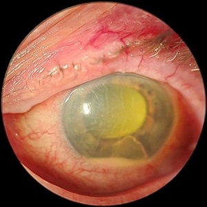 Acute Endophthalmitis
Nov 14 2023 by Virginia Gebhart
85 year old female with acute endophthalmitis 14 days s/p IVEylea injection. 3+ injection and descemets folds, contracted fibron, 3mm Hypopyon, and 3+ cell. No view posteriorly, vision CF. Visual prognosis unknown at this time
Photographer: Virginia Gebhart
Condition/keywords: endophthalmitis
-
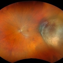
 Geographic Atrophy
Nov 16 2023 by Virginia Gebhart
67 year old female with Neovascular AMD with inactive CNV. Extensive geographic atrophy with minimal foveal sparing. Extensive ectopic CNV just superiorly to ON remains inactive. Discussed with pt treating with Syfovre to slow down GA progression
Photographer: Virginia Gebhart
Imaging device: Optos
Condition/keywords: advanced geographic atrophy, age-related macular degeneration (AMD), dry age-related macular degeneration (dry AMD), geographic atrophy
-
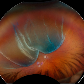
 New Retinal Detachment 6w s/p RD repair
Nov 16 2023 by Virginia Gebhart
13 year old male presented with new blind spot 6 weeks s/p RD repair with cryo/scleral buckle/prophylaxis laser with gas bubble. New RD involving the macula, posterior to scleral buckle, secondary to PVD. Small gas bubble remaining. Pt was brought back to OR for repeat PPV and silicone oil repair
Photographer: Virginia Gebhart
Imaging device: Optos
Condition/keywords: gas bubble, Retinal Detachment, retinal detachment of the macula, scleral buckle
-
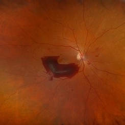
 Fibrotic granuloma vs. Pseudoduplication of the Optic Disc
Nov 29 2023 by Virginia Gebhart
74 year-old female with presumed fibrotic granuloma. Previously diagnosed as pseudoduplication of the optic disc by general ophthalmologist. OCT showed elevation in the RPE, more consistent with granuloma. Pt has been aware for many years, asymptomatic. Will observe.
Photographer: Virginia Gebhart
Imaging device: Topcon 50DX
Condition/keywords: fibrotic scar, granuloma, Pseudoduplication of optic disc
-
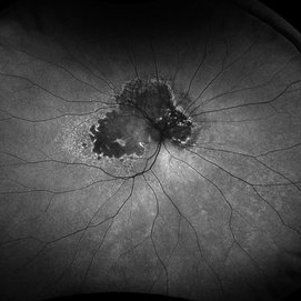
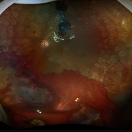
 Layer Cake; Sub-retinal, Pre-retinal, Vitreous Hemorrhages
Dec 5 2023 by Virginia Gebhart
73 year old female with sub-retinal, pre-retinal, and vitreous hemorrhages all in OD. Will consider sx if blood does not clear on its own. Vision 20/40
Photographer: Virginia Gebhart
Imaging device: Topcon
Condition/keywords: pre-retinal hemorrhage, retinal macroaneurysm, subretinal hemorrhage, subretinal blood, vitreous hemorrhage
-
 Mac off Retinal Detachment with Horseshoe Tear
Dec 5 2023 by Virginia Gebhart
68 year old male presented with HM vision in OD. Near total detachment with multiple breaks. Scheduled PPV with GFE. Visual prognosis guarded
Photographer: Virginia Gebhart
Imaging device: Topcon
Condition/keywords: Retinal Detachment, retinal detachment of the macula, Retinal Detachment with multiple breaks
-
 Familial Dominant Drusen
Dec 5 2023 by Virginia Gebhart
50 year old female with presumed Familial Dominant Drusen OU. Asymptomatic, will observe
Photographer: Virginia Gebhart
Imaging device: Topcon
Condition/keywords: Drusen, FAMILIAL DOMINANT DRUSEN, macular drusen
-
 Regressed Choroidal Metastasis
Dec 6 2023 by Virginia Gebhart
60 year old female with regressing metastasis along STA. Tumor has continued to regress s/p radiation in Feb 2023 and Tagrisso. VA 20/20. No elevation or SRF on most recent ultrasound
Photographer: Virginia Gebhart
Imaging device: Topcon
Condition/keywords: choroidal metastasis, choroidal tumor
-
 Suspicious Choroidal Nevus
Dec 6 2023 by Virginia Gebhart
82 year old male with suspicious choroidal nevus and ERM. Remains unchanged since first visit 6 mos ago.
Photographer: Virginia Gebhart
Imaging device: Topcon
Condition/keywords: choroidal nevus
-
 New Choroidal Melanoma vs Metastasis
Dec 6 2023 by Virginia Gebhart
72 year old male with possible new choroidal melanoma vs metastatic melanoma. Dome-shaped amelanotic lesion involving the fovea. Lesion was discovered during a problem visit due to sudden decreased VA (20/150). Most recent CT scan shows concern for primary lung cancer
Photographer: Virginia Gebhart
Imaging device: Topcon
Condition/keywords: amelanotic melanoma, melanoma, metastatic lesion
-
 Benign Nevus
Dec 6 2023 by Virginia Gebhart
71 year old female with benign choroidal nevus with fibrotic PED
Photographer: Virginia Gebhart
Imaging device: Topcon
Condition/keywords: choroidal nevus, fibrotic neovascularization, nevus
-
 Suspicious Choroidal Nevus
Dec 6 2023 by Virginia Gebhart
55 year old female with suspicious pigmented choroidal nevus. 3 high risk features present. Ultrasound shows SRF and high internal reflectivity. Will observe closely
Photographer: Virginia Gebhart
Imaging device: Topcon
Condition/keywords: choroidal nevus, orange pigment
-
 Choroidal Metastasis
Dec 6 2023 by Virginia Gebhart
60 year old female with totally regressed tumor in temporal macula s/p external beam radiation and chemo. Pt diagnosed with stage IV metastatic lung cancer.
Photographer: Virginia Gebhart
Imaging device: Optos
Condition/keywords: choroidal metastasis, choroidal tumor
-
 Tilted IOL
Dec 7 2023 by Virginia Gebhart
61 year old female with inferiorly tilted IOL. UBM shows haptic rubbing against the iris causing transillumination defect from 5 to 6 o'clock.
Photographer: Virginia Gebhart
Condition/keywords: lens dislocation, transillumination
-
 The Nightmare Before Christmas
Dec 12 2023 by Virginia Gebhart
78 year old female with intermediate stage dry AMD. Large soft drusen in macula as well as drusenoid pigment epithelial detachment.
Photographer: Virginia Gebhart
Imaging device: Optos
Condition/keywords: drusenoid PED, macular drusen
-
 Macular Dystrophy vs Myopic Degeneration
Dec 22 2023 by Virginia Gebhart
35 year old female with myopic degeneration (-18.00 OU). BCVA 20/100 OU. RPE atrophy present in both eyes, but no significant chorioretinal atrophy. OCT not consistent with degenerative myopia due to dome shape appearance rather than posterior bowing. Possible macular dystrophy over degeneration. Will observe
Photographer: Virginia Gebhart
Imaging device: Topcon
Condition/keywords: Macular Dystrophy, myopic degeneration
-
 PEHCR
PEHCR
Jan 4 2024 by Virginia Gebhart
86 year old male with partially oxidized choroidal hemorrhage and CME. Previous FA shows blocking defect temporally, most likely a choroidal hemorrhage with SRH and late leakage. Continued improvement with 8 week intervals of Eylea. VA 20/60 Previous RD repair with scleral buckle and cryo in 1980's
Photographer: Virginia Gebhart
Imaging device: Optos California
Condition/keywords: chorioretinopathy, choroidal hemorrhage, cystoid macular edema (CME), peripheral exudative hemorrhagic chorioretinopathy (PEHCR)
-
 New Choroidal Melanoma
Jan 4 2024 by Virginia Gebhart
77 year old male with a bilobed pigmented mass with exudative RD, and trace inflammation present in AV consistent with choroidal melanoma. Mass extends into ciliary body. Pt scheduled for MRI prior to plaque radiation to rule out metastasis.
Photographer: Virginia Gebhart
Imaging device: Optos California
Condition/keywords: ciliary body melanoma, exudative retinal detachment
-
 Idiopathic Peripapillary CNV
Jan 4 2024 by Virginia Gebhart
13 year old female with inactive CNV. Increased pigment 360 at 1 year follow up. No inflammation or SRF, pt remains asymptomatic
Photographer: Virginia Gebhart
Imaging device: Optos California
Condition/keywords: choroidal neovascularization (CNV), peripapillary choroidal neovascularization (PPCNVM)
-
 Choroidal Melanoma
Jan 4 2024 by Virginia Gebhart
57 year old female with new choroidal melanoma. Early hyperfluorescence with vascularity and minimal late leakage on FA.
Photographer: Virginia Gebhart
Imaging device: Optos California
Condition/keywords: FA, FA early phase, fluorescein angiogram (FA), Fluorescein angiography
-
 Preretinal Fibrosis
Jan 12 2024 by Virginia Gebhart
53 year old diabetic male with significant persistent ERM due to fibrotic NV superiorly. Possibly developing a tractional MH. Vitreous Hemorrhage secondary to traction on the fibrosis
Photographer: Virginia Gebhart
Imaging device: Topcon 50DX
Condition/keywords: epiretinal membrane, ERM, fibrosis, macular pseudohole, neovascularization (NV)
-
 Retinitis Pigmentosa
Jan 25 2024 by Virginia Gebhart
58 year old female diagnosed at age 13. Vision 20/100
Photographer: Virginia Gebhart
Imaging device: Topcon
Condition/keywords: retinitis pigmentosa, retinitis pigmentosa (RP) dystrophy
-
 Hemorrhagic Pigment Epithelial Detachment
Jan 25 2024 by Virginia Gebhart
64 year old male with persistent hemorrhagic PED with oxidized SRH involving the central macula. Continued improvement with 12 week intervals of Eylea. BCVA 20/80
Photographer: Virginia Gebhart
Imaging device: Topcon
Condition/keywords: neovascular age-related macular degeneration (AMD), pigment epithelial detachment (PED)
-
 Dry AMD
Jan 25 2024 by Virginia Gebhart
79 year old female with intermediate dry AMD. Small area of geographic atrophy superior, large drusen and stippled RPE changes. BCVA 20/40
Photographer: Virginia Gebhart
Imaging device: Topcon
Condition/keywords: age-related macular degeneration (AMD), dry age-related macular degeneration (dry AMD), geographic atrophy
-
 Retinal Detachment with PVR
Jan 25 2024 by Virginia Gebhart
62 year old female with history of RD repair with scleral buckle 30 years ago. Low scleral buckle with cryo scar, attached anteriorly. New RD repaired with gas bubble
Photographer: Virginia Gebhart
Imaging device: Topcon
Condition/keywords: proliferative vitreoretinopathy (PVR), Retinal Detachment
-
 RPE Rip
Jan 25 2024 by Virginia Gebhart
69 year old female with Neovascular AMD. New RPE rip and increased IRF on OCT 10 weeks s/p Eylea injection. Switched to Vabysmo to extend intervals
Photographer: Virginia Gebhart
Imaging device: Topcon
Condition/keywords: neovascular age-related macular degeneration (AMD)
-
Persistent TRD with Sub-hyaloid Hemorrhage
Jan 25 2024 by Virginia Gebhart
Progression of TRD in 46 year old male from March 2023 to now. Pt complains of worsening vision over past 3 weeks. Surgery recommended to remove scar tissue.
Condition/keywords: Diabetic Tractional Retinal Detachment involving the Macula, Proliferative Diabetic retinopathy, Sub-Hyaloid Hemorrhage, tractional retinal detachment, vitreous hemorrhage
-
 Iris Melanoma
Feb 1 2024 by Virginia Gebhart
90 year old female with elevated pigmented lesion, amelanotic portion extending toward the angle, questionable vascularity on UBM.
Photographer: Virginia Gebhart
Imaging device: Samsung Galaxy Z Flip
Condition/keywords: iris lesion, iris melanoma
-
 Melanocytosis
Feb 6 2024 by Virginia Gebhart
64 year old male with benign appearing choroidal pigmentation consistent with melanocytosis.
Photographer: Virginia Gebhart
Imaging device: Topcon TRC 50DX
Condition/keywords: choroidal melanocytosis
-
 Diabetic Macular Edema
Feb 7 2024 by Virginia Gebhart
FA of 70 year old male with diabetic macular edema. FA shows early hyper-fluorescence with late leakage and capillary dropout in the temporal macula. Focal laser performed.
Photographer: Virginia Gebhart
Imaging device: Optos California
Condition/keywords: capillary dropouts, fluorescein angiogram (FA), macular edema
-
 Treated Choroidal Melanoma
Feb 7 2024 by Virginia Gebhart
40 year old male with a stable, flat, treated tumor s/p brachytherapy in 2018. Persistent CME secondary to radiation retinopathy.
Photographer: Virginia Gebhart
Imaging device: Optos California
Condition/keywords: melanoma, radiation retinopathy
-
 Suspicious Nevus
Feb 14 2024 by Virginia Gebhart
61 year old female with a suspicious choroidal nevus involving the optic nerve head. Patient asymptomatic, will continue to observe.
Photographer: Virginia Gebhart
Imaging device: Topcon TRC 50DX
Condition/keywords: choroidal nevus, nevus
-
 Diffuse Chorioretinal Atrophy
Feb 21 2024 by Virginia Gebhart
61 year male with myopic degeneration and diffuse chorioretinal atrophy. BCVA 20/200.
Photographer: Virginia Gebhart
Imaging device: Topcon TRC 50DX
Condition/keywords: chorioretinal atrophy, myopic degeneration
-
 Optic Nerve Pit
Feb 21 2024 by Virginia Gebhart
65 year old female with optic nerve pit. Asymptomatic, continued observation.
Photographer: Virginia Gebhart
Imaging device: Topcon TRC 50DX
Condition/keywords: congenital optic nerve pit, Optic nerve pit
-
 Hidden Mickey / Toxo Scar
Feb 29 2024 by Virginia Gebhart
87 year old female with inactive toxoplasmosis chorioretinitis inferior. Stable regressed malignant neoplasm of choroid superior (s/p brachytherapy 2011).
Photographer: Virginia Gebhart
Imaging device: Optos California
Condition/keywords: inactive toxoplasmosis, toxo chorioretinitis, toxoplasmosis chorioretinitis
-
 Choroidal Melanoma
Mar 1 2024 by Virginia Gebhart
52 year old female at first visit July 2023 vs 7 months s/p brachytherapy. SRF in macula has resolved, trace fluid on posterior edge of collapsing collar button.
Photographer: Virginia Gebhart
Imaging device: Optos California
Condition/keywords: brachytherapy, Choroidal melanoma, collar button
-
 PDR
PDR
Mar 15 2024 by Virginia Gebhart
FA of 59 year old female with proliferative diabetic retinopathy.
Photographer: Virginia Gebhart
Imaging device: Optos California
Condition/keywords: Diabetic Retinopathy, neovascularization of the disc (NVD), NVE, proliferative diabetic retinopathy (PDR)
-
 Retinal Detachment
Mar 28 2024 by Virginia Gebhart
68 year male with chronic appearing retinal detachment with subretinal bands and subretinal fibrosis. Demarcation line present, SRF splits the fovea on OCT.
Photographer: Virginia Gebhart
Imaging device: Optos California
Condition/keywords: chronic retinal detachment, Retinal Detachment
-
 Benign Lobular Inner Nuclear Layer Proliferations (BLIP)
Apr 15 2024 by Virginia Gebhart
29 year old male with multiple flat CHRPE lesions, genetic testing negative for ACP genes associated with Gardner syndrome. Multiple intraretinal amelanotic lesions consistent with Benign Lobular Inner Nuclear Layer Proliferations (BLIP) of the retina
Photographer: Virginia Gebhart
Imaging device: Topcon
Condition/keywords: BLIP Benign Lobular Inner Nuclear Layer Proliferations, CHRPE, congenital hypertrophy of the retinal pigment epithelium (CHRPE)
-
 Peripheral Retinal Degeneration (L-ORD)
Apr 17 2024 by Virginia Gebhart
92 year old female with bilateral patchy, sharply demarcated circular areas of chorioretinal atrophy with hyperpigmented margins in the mid to far periphery. Labs showed normal plasma ornithine levels ruling out generalized gyrate atrophy. Also intermediate uveitis and CMD/CME. FTA-ABS, Quant gold, and HLA-A29 labs all negative.
Photographer: Virginia Gebhart
Imaging device: Optos California
Condition/keywords: cystoid macular degeneration, cystoid macular edema (CME), FA, Fluorescein angiography, peripheral retinal degeneration
-
 Sturge-Weber Syndrome
May 8 2024 by Virginia Gebhart
13 year old female with diffuse hemangioma consistent with Sturge-Weber Syndrome. Hx of POAG
Photographer: Virginia Gebhart
Imaging device: Optos California
Condition/keywords: diffuse choroidal hemangioma, Hemangioma, Sturge-Weber syndrome
-
 Suspicious Choroidal Nevus
May 8 2024 by Virginia Gebhart
13 year old female with suspicious appearing choroidal nevus. High risk features present, adjacent to optic nerve, questionable orange pigment, SRF. No significant elevation on ultrasound. Will follow up with serial exams.
Photographer: Virginia Gebhart
Imaging device: Optos California
Condition/keywords: choroidal nevus, nevus
-
 Choroidal Folds s/p External Beam Radiation
May 15 2024 by Virginia Gebhart
74 year old female with choroidal folds s/p external beam radiation 11/2023. Choroidal infiltration and resolved SRF most likely secondary to Chronic Lymphocytic Leukemia. Pt remains asymptomatic.
Photographer: Virginia Gebhart
Imaging device: Topcon 50DX
Condition/keywords: after proton beam irradiation, choroidal folds
-
 Choroidal Osteoma
May 30 2024 by Virginia Gebhart
33 year old female with regressed osteoma OS s/p focal laser, TTT and PDT (first treatment in 2013). Vision 20/20, pt remains asymptomatic.
Photographer: Virginia Gebhart
Imaging device: Topcon 50DX
Condition/keywords: choroidal osteoma
-
 Retinal Detachment with Single Break
May 30 2024 by Virginia Gebhart
73 year old female with new mac-off retinal detachment with single horseshoe tear. Vision CF@3ft. Repaired in OR with gas bubble and laser. Guarded prognosis
Photographer: Virginia Gebhart
Imaging device: Optos California
Condition/keywords: retinal detachment of the macula
-
 Choroidal Osteoma
Jun 13 2024 by Virginia Gebhart
20 year old female with choroidal osteoma. Stable s/p PDT x3 and focal laser x 2, no obvious progression on last exam. Monitoring closely. Vision 20/30.
Photographer: Virginia Gebhart
Imaging device: Topcon 50 DX
Condition/keywords: barrier laser, choroidal osteoma, PDT
-
 Hollenhorst Plaque
Jun 25 2024 by Virginia Gebhart
75 year female with complaint of shadow in the bottom of her vision for many years. Hollenhorst plaque on superior pole of the disc and sclerotic superotemporal arteriole. Also DBHs superiorly most likely due to combined BRAO/BRVO.
Photographer: Virginia Gebhart
Imaging device: Topcon 50DX
Condition/keywords: branch retinal artery occlusion (BRAO), branch retinal vein occlusion (BRVO), hollenhorst plaque, sclerotic arteriole
-
 Focal Chorioretinitis
Jul 11 2024 by Virginia Gebhart
67 year old female with punched-out CR scars. Hx of laser 3x for apparent peripapillary CNV. ESR, CRP, toxo, IgG/IgM all "normal." Bartonella, quant gold, and FTA-ABS ordered given possibility of neuroretinitis. Vision CF
Photographer: Virginia Gebhart
Imaging device: Optos California
Condition/keywords: FA, fluorescein angiogram (FA), FLUORESCEIN ANGIOGRAPHY, focal chorioretinitis, optic neuritis
-
 Twinkle Twinkle
Aug 5 2024 by Virginia Gebhart
65 year old male with mac-off retinal detachment with 360 folds and horseshoe tear.
Photographer: Virginia Gebhart
Imaging device: Optos California
Condition/keywords: macula off retinal detachment, RD, Retinal Detachment
-
 Choroidal Melanoma
Aug 8 2024 by Virginia Gebhart
88 year old male with new bilobed choroidal melanoma. Pt scheduled for brachytherapy
Photographer: Virginia Gebhart
Imaging device: Optos California
Condition/keywords: melanoma
-
 Birdshot Retinochoroiditis
Aug 8 2024 by Virginia Gebhart
ICG angiogram of 45 year old male with Birdshot Retinochoroiditis. Has been improving on Humira and Methotrexate.
Photographer: Virginia Gebhart
Imaging device: Optos California
Condition/keywords: birdshot, indocyanine green (ICG) angiography
-
 Suspicious Nevus / CSR
Aug 8 2024 by Virginia Gebhart
Fluorescein angiogram of 54 year old male with a suspicious appearing choroidal nevus and central serous retinopathy. Will monitor closely and follow up with serial exams.
Photographer: Virginia Gebhart
Imaging device: Optos California
Condition/keywords: central serous retinopathy (CSR), choroidal nevus, fluorescein angiogram (FA), FLUORESCEIN ANGIOGRAPHY, nevus
-
 Fundus Flavimaculatus
Aug 12 2024 by Virginia Gebhart
77 year old female with symmetrical retinal flecks consistent with hereditary dystrophy. Unable to complete genetic testing today, will consider in the future. Pt Asymptomatic, Dcc 20/20 OU
Photographer: Virginia Gebhart
Imaging device: Optos California
Condition/keywords: fundus flavimaculatus
-
 Diabetic Tractional Retinal Detachment 1 week s/p SO fill
Aug 14 2024 by Virginia Gebhart
21 year old male 1 week s/p PPV/laser/STR/SO. Eye is stable, PRHs inferior and superior, possible traction from PRH/membrane. Will observe and let clot liquify, will consider scleral buckle if no improvement
Photographer: Virginia Gebhart
Imaging device: Optos California
Condition/keywords: Diabetic Tractional Detachment, retinal detachment of the macula, silicone oil
-
 Branch Retinal Vein Occlusion
Aug 22 2024 by Virginia Gebhart
Fluorescein angiogram of branch retinal vein occlusion in 75 year old female. Scattered microaneurysms with late CME and persistent SRF. Pt will consider laser treatment but is hesitant for injections at this time due to possible side effects.
Photographer: Virginia Gebhart
Imaging device: Optos California
Condition/keywords: branch retinal vein occlusion (BRVO), BRVO, cystoid macular edema (CME), FA, FA late phase, fluorescein angiogram (FA), macular edema, microaneurysms, retinal microaneurysms
-
 Pre-Retinal Hemorrhage
Aug 22 2024 by Virginia Gebhart
51 year old female with moderate proliferative diabetic retinopathy, DME, as well as pre-retinal hemorrhage and likely NVE. Pt given Avastin in office and will return for PRP.
Photographer: Virginia Gebhart
Imaging device: Optos California
Condition/keywords: diabetic macular edema, macular edema, PDR with NVE (periphery), pre-retinal hemorrhage, proliferative diabetic retinopathy (PDR)
-
 New RD with Multiple Breaks
Aug 26 2024 by Virginia Gebhart
59 year old male with superior, bullous, mac off RD with multiple breaks. Pt scheduled for PPV, laser, GFE.
Photographer: Virginia Gebhart
Imaging device: Optos California
Condition/keywords: retinal detachment of the macula, retinal tear, retinal tear with detachment
-
 Repaired Retinal Detachment
Aug 26 2024 by Virginia Gebhart
13 year old male 2 weeks s/p silicone oil placement and lensectomy. (Previous scleral buckle placement in 2023). Retina remains attached on the buckle under oil.
Photographer: Virginia Gebhart
Imaging device: Optos California
Condition/keywords: RD, Retina detachment, Retinal detachment under Silicon Oil, scleral buckle
-
 Sturge-Weber Syndrome
Sep 4 2024 by Virginia Gebhart
13 year old female with hemangioma of the retina and history of Sturge-Weber syndrome. Slightly larger compared to photos and ultrasound from 4 months ago. SRF inferior on ultrasound and clinical exam. Pt and mom will consider brachytherapy.
Photographer: Virginia Gebhart, Retina Consultants of Carolina
Imaging device: Optos California
Condition/keywords: diffuse choroidal hemangioma, Hemangioma, Sturge-Weber syndrome
-
 Choroidal Melanoma
Sep 5 2024 by Virginia Gebhart
63 year old female with new choroidal melanoma. Pt scheduled for brachytherapy.
Photographer: Virginia Gebhart, Retina Consultants of Carolina
Imaging device: Optos California
Condition/keywords: choroidal melanoma, melanoma
-
 Horseshoe Tear with Vitreous Hemorrhage
Sep 19 2024 by Virginia Gebhart
New horseshoe tear without detachment in 64 year old male. Vitreous hemorrhage secondary to HST. Prophylactic laser performed to seal tear
Photographer: Virginia Gebhart, Retina Consultants of Carolina
Imaging device: Optos California
Condition/keywords: retinal tear, vitreous hemorrhage
-
 Uveal Effusion Syndrome
Sep 19 2024 by Virginia Gebhart
61 year old female with idiopathic uveal effusion syndrome. 360 degrees of choroidal thickening, especially anterior with exudative fluid inferior. Mild vitritis present. Unable to gain venous access for FA, ultrasound and UBM performed which confirm choroidal and ciliary body thickening. Pt sent for inflammatory work up including MRI of brain and orbits. Treatment pending results.
Photographer: Virginia Gebhart, Retina Consultants of Carolina
Imaging device: Optos California
Condition/keywords: idiopathic uveal effusion syndrome, uveal effusion
-
 POHS/Schlaegel Lines
Sep 19 2024 by Virginia Gebhart
46 year old female with h/o Histoplasmosis. Multiple punched out chorioretinal scars with Schlaegel lines. No evidence of CNV or active inflammation. VA 20/20
Photographer: Virginia Gebhart, Retina Consultants of Carolina
Imaging device: Optos California
Condition/keywords: chorioretinal scar, histoplasmosis, presumed ocular histoplasmosis syndrome (POHS)
-
 Look With Your Heart
Sep 20 2024 by Virginia Gebhart
FA of 65 year old male with exudative AMD superior to a chorioretinal defect in the nasal macula. FA shows classic CNV with late leakage. Treated with IVA, will consider PDT if no improvement.
Photographer: Virginia Gebhart, Retina Consultants of Carolina
Imaging device: Optos California
Condition/keywords: choroidal neovascularization (CNV), exudative age-related macular degeneration, FA early phase
-
 New Iris Melanoma
Oct 10 2024 by Virginia Gebhart
56 year old male with new amelanotic melanoma emanating from the ciliary body through the posterior iris epithelium. CT scan showed no evidence of metastatic disease. Pt scheduled for radioactive plaque and tumor biopsy
Photographer: Virginia Gebhart, Retina Consultants of Carolina
Imaging device: Samsung Galaxy
Condition/keywords: amelanotic melanoma, iris melanoma
-
 IOFB
IOFB
Oct 11 2024 by Virginia Gebhart
55 year old male s/p RD repair with SB and SO in Mexico June 2023. Questionable foreign body inferior vitreous base. Pt asymptomatic, had no previous knowledge of IOFB.
Photographer: Virginia Gebhart, Retina Consultants of Carolina
Imaging device: Optos California
Condition/keywords: intraocular foreign body, IOFB, scleral buckle
-
 Suspicious Lesion 18 Years s/p Iris Resection
Oct 15 2024 by Virginia Gebhart
85 year old female with small pigmented lesion present s/p sectoral iridectomy in 2006. Lesion is suspicious for recurrence of melanoma after 18 years. Stable compared to previous exam in March 2024, unclear if this is a new lesion or has been present for an extended time. Will monitor closely.
Photographer: Virginia Gebhart, Retina Consultants of Carolina
Imaging device: Samsung Galaxy
Condition/keywords: iris melanoma, melanoma
-
 Retinitis Pigmentosa
Oct 16 2024 by Virginia Gebhart
74 year old female with bone spicule pigmentation associated with Retinitis Pigmentosa. Pt diagnosed at age 53, relatively asymptomatic prior to diagnosis. Pt reports gradual vision loss over 10+ years. BCVA 20/40
Photographer: Virginia Gebhart, Retina Consultants of Carolina
Imaging device: Optos California
Condition/keywords: bone spicule, retinitis pigmentosa, retinitis pigmentosa (RP) dystrophy
-
 New Choroidal Melanoma with Exudative Detachment
Oct 16 2024 by Virginia Gebhart
56 year old male with a large pigmented tumor with an exudative detachment inferior and shallow fluid through the macula. Pt states they have been having symptoms for over a year. Scheduled for brachytherapy.
Photographer: Virginia Gebhart, Retina Consultants of Carolina
Imaging device: Optos California
Condition/keywords: Choroidal melanoma, exudative detachment, melanoma
-
 Stargardt's Disease
Oct 23 2024 by Virginia Gebhart
62 year old female with bullseye RPE changes and flecks, mottled FAF, and silent choroid on FA consistent with late onset Stargardt's Disease. Pt is asymptomatic with 20/20 vision OU at this time
Photographer: Virginia Gebhart, Retina Consultants of Carolina
Imaging device: Optos California
Condition/keywords: Stargardt disease, Stargardts Disease
-
 Ciliary Body Melanoma
Nov 2 2024 by Virginia Gebhart
53 year old male with a large mass behind the lens as well as prominent scleral vessels. Clinical exam and ultrasound findings consistent with melanoma. Pt will be scheduled for enucleation pending CT scan results. Edit: Sadly patient has canceled all appointments and has requested no further contact
Photographer: Virginia Gebhart, Retina Consultants of Carolina
Imaging device: Optos California
Condition/keywords: ciliary body mass, ciliary body melanoma, ciliary body tumor
-
 Recurrent Retinal Detachment with Single Break
Nov 2 2024 by Virginia Gebhart
84 year old male with recurrent detachment s/p PPV/RD repair 2 weeks ago. Retinotomy is opened and appears to be the source of the fluid. Pt scheduled for emergency repair with scleral buckle.
Photographer: Virginia Gebhart
Imaging device: Optos California
Condition/keywords: gas bubble, retinal detachment, retinotomy
-
 New Choroidal Melanoma
Nov 7 2024 by Virginia Gebhart
83 year old female with new choroidal melanoma. Diffuse, flat tumor with orange pigment, SRF and located adjacent to ON. Pt sleeps on left side causing fluid to pool in/around the macula. Pt scheduled for brachytherapy pending CT scan results.
Photographer: Virginia Gebhart, Retina Consultants of Carolina
Imaging device: Optos California
-
 Best Disease
Nov 7 2024 by Virginia Gebhart
Fluorescein angiogram of 49 year female with Best Disease. Genetic testing done in 2000 confirms Best Disease and also possible Stargardts mutation. Characteristic bullseye maculopathy with surrounding yellowish flecks are present in both eyes.
Photographer: Virginia Gebhart, Retina Consultants of Carolina
Imaging device: Optos California
Condition/keywords: Best disease, fluorescein angiogram (FA)
-
 Retinal Telangiectasis
Nov 13 2024 by Virginia Gebhart
42 year old female with telangiectatic vessels and vascular sheathing. FA showed mild leakage with areas of peripheral non-perfusion. Pt is asymptomatic without inflammation in the vitreous. No history of systemic inflammatory disease. Will observe.
Photographer: Virginia Gebhart, Retina Consultants of Carolina
Imaging device: Optos California
Condition/keywords: retinal telangiectasia, telangiectatic vessels, vascular sheathing of retina
-
 PDR/Vitreous Hemorrhage
Nov 20 2024 by Virginia Gebhart
76 year old male with new proliferative diabetic retinopathy. NVD and VH on exam. Pt treated with IVEylea, will consider PRP in the near future.
Photographer: Virginia Gebhart, Retina Consultants of Carolina
Imaging device: Optos California
Condition/keywords: PDR, proliferative diabetic retinopathy (PDR), VH, vitreous hemorrhage
-
 Pigmentary Degeneration of Retina (Secondary to Elmiron)
Nov 27 2024 by Virginia Gebhart
77 year old female with advanced geographic atrophy after years of Elmiron use (stopped in 2018). Serial exams show continued progression of GA. Central vision limited, vision remains stable and patient does not report noticing any changes.
Photographer: Virginia Gebhart, Retina Consultants of Carolina
Imaging device: Optos California
Condition/keywords: geographic atrophy, secondary pigmentary degeneration, toxic maculopathy
-
 Rosai-Dorfman Disease
Dec 4 2024 by Virginia Gebhart
72 year old female with temporal limbal lesion that extends onto the cornea from 10:00 - 8:00 encroaching on visual axis. Possible lymphomatous process. Will refer to Emory.
Photographer: Dr Chris Bergstrom MD, OD
Condition/keywords: corneal scars and opacities, Rosai-Dorfman Disease, Subconjunctival mass
-
 Reactive Retinal Astrocytic Tumor
Dec 6 2024 by Virginia Gebhart
27 year old female self-referred for continued follow-up care of hemangioma of retina. Previous genetic testing negative for Von Hippel-Lindau. Pt recently diagnosed with Ehlers-Danlos Arthroclasia, most likely reactive retinal astrocytic tumor. Tumor is stable and surrounded by good laser barricade, will continue to observe.
Photographer: Virginia Gebhart, Retina Consultants of Carolina
Imaging device: Optos California
Condition/keywords: feeder vessel, hemangioma, RRAT
-
 Repaired Retinal Detachment with Multiple Breaks
Dec 9 2024 by Virginia Gebhart
FAF in 25 year old female of repaired retinal detachment 1.5 year s/p scleral buckle/cryo. Pt had been having symptoms for over a year, inferior demarcation line from retinal fluid that was present. Retina remains flat and attached under buckle. Treated lattice inferiorly, no new holes or tears. VA 20/20
Photographer: Virginia Gebhart, Retina Consultants of Carolina
Imaging device: Optos California
Condition/keywords: autofluorescence imaging, cryotherapy, demarcation line, lattice degeneration, scleral buckle
-
 Myopic Degeneration
Dec 9 2024 by Virginia Gebhart
67 year old female with myopic degeneration. Posterior staphylomas are stable. VA limited by extensive chorioretinal atrophy. BCVA 20/160 (ecc)
Photographer: Virginia Gebhart, Retina Consultants of Carolina
Imaging device: Optos California
Condition/keywords: chorioretinal atrophy, myopic degeneration, staphyloma
-
 MEWDS
MEWDS
Dec 11 2024 by Virginia Gebhart
28 year old female with new Multiple Evanescent White Dot Syndrome. Patient reports gray spot in vision, OCT shows RPE disruption centrally but no edema. FA shows early hyperfluorescent punctate spots throughout the posterior pole, but no leakage. Normal findings OD. Will observe for now
Photographer: Virginia Gebhart, Retina Consultants of Carolina
Imaging device: Optos California
Condition/keywords: FA, fluorescein angiogram (FA), multiple evanescent white dot syndrome (MEWDS)
-
 Inactive Chorioretinal Scars
Dec 11 2024 by Virginia Gebhart
30 year old female with chorioretinal and macula scars. Appears post-infectious, most likely toxoplasmic. No active inflammatory changes or choroidal neovascularization. Will continue to monitor. Central vision limited by macula scar, BCVA 20/100
Photographer: Virginia Gebhart, Retina Consultants of Carolina
Imaging device: Optos California
Condition/keywords: chorioretinal scar, inactive toxoplasmosis
-
 Guardian Angel
Dec 11 2024 by Virginia Gebhart
48 year old female 3 months s/p brachytherapy for choroidal melanoma. Persistent subretinal and increased subfoveal fluid. Will observe for now, will consider Ozurdex if no improvement. BCVA 20/80
Photographer: Virginia Gebhart, Retina Consultants of Carolina
Imaging device: Optos California
Condition/keywords: brachytherapy, demarcation line, fundus autofluorescence (FAF), serous detachment, subretinal fluid
-
 Snowbank in Intermediate Uveitis
Dec 12 2024 by Virginia Gebhart
25 year old female with intermediate uveitis. Improved inflammation after 40mg prednisone taper. Pt has multiple autoimmune diseases, will order MRI to r/o demyelinating disease.
Photographer: Virginia Gebhart, Retina Consultants of Carolina
Imaging device: Optos California
Condition/keywords: intermediate uveitis, snowballs, snowbank, uveitis
-
 Coloboma in a Unicameral Eye
Dec 20 2024 by Virginia Gebhart
59 year old female with choroidal coloboma extending into iris. Pt had PC IOL placed in 2016, removed in Aug due to suspected UGH syndrome. Lens haptics were oriented vertically causing haptic to chafe iris superiorly. Most likely etiology was loss of inferior zonules from coloboma. Pt remains aphakic.
Photographer: Virginia Gebhart, Retina Consultants of Carolina
Imaging device: Optos California
Condition/keywords: choroidal coloboma, coloboma
-
 Repaired Retinal Detachment with Grade C PVR
Dec 23 2024 by Virginia Gebhart
61 year old male 1 day s/p retinectomy/SO exchange. Retina is attached under SO with good laser to retinectomy edge.
Photographer: Virginia Gebhart, Retina Consultants of Carolina
Imaging device: Optos California
Condition/keywords: gas bubble, proliferative vitreoretinopathy (PVR), retinectomy, silicone oil, total retinal detachment
-
 Giant Retinal Tear with Detachment
Jan 3 2025 by Virginia Gebhart
67 year old male with mac-on RD with giant tear. Pt scheduled for sx
Photographer: Virginia Gebhart
Imaging device: Optos California
Condition/keywords: giant retinal tear, retinal tear with detachment
-
 New Choroidal Melanoma
Jan 3 2025 by Virginia Gebhart
22 year old male referred for 2nd opinion on large choroidal mass with subretinal fluid. Clinical exam and ultrasound consistent with choroidal melanoma. CT scan of orbits showed possible inflammation involving orbital fat. Pt has been on oral prednisone for 1 week, inflammation has not responded. Referred to Emory for 2nd opinion on treatment
Photographer: Virginia Gebhart
Imaging device: Optos California
Condition/keywords: melanoma
-
 Melanocytoma of Optic Disc
Jan 13 2025 by Virginia Gebhart
25 year-old female referred for melanocytoma of optic disc. Lesion is benign, no treatment necessary. Pt asymptomatic.
Photographer: Virginia Gebhart, Retina Consultants of Carolina
Imaging device: Topcon 50DX
Condition/keywords: benign melanocytoma, melanocytoma, optic disc melanocytoma
-
 Suspicious Nevus
Jan 15 2025 by Virginia Gebhart
14 year female with suspicious nevus located adjacent to the optic nerve. Questionable orange pigment present and worsening SRF compared to previous photos/OCT. RPE atrophy also present from previous fluid. No elevation. Will continue observation. BCVA 20/25
Photographer: Virginia Gebhart, Retina Consultants of Carolina
Imaging device: Topcon 50DX
Condition/keywords: choroidal nevus, nevus
-
 Elmiron Toxicity
Jan 15 2025 by Virginia Gebhart
54 year old female with pigmentary degeneration secondary to Elmiron. Stippled RPE maculopathy has lightly progressed with stable vision compared to previous visits. BCVA 20/200 OU. Pt reports taking Elmiron from 2010 to 2019.
Photographer: Virginia Gebhart
Imaging device: Optos California
Condition/keywords: autofluorescence imaging, Maculopathy, secondary pigmentary degeneration
-
 Retinitis Pigmentosa
Jan 15 2025 by Virginia Gebhart
52 year old male with advanced RP OU. BCVA HM OD, LP OS. Referred to genetic specialist per pt request to discuss gene therapy.
Photographer: Virginia Gebhart, Retina Consultants of Carolina
Imaging device: Optos California
Condition/keywords: bone spicule, retinitis pigmentosa, retinitis pigmentosa (RP) dystrophy
-
 CRVO
CRVO
Jan 15 2025 by Virginia Gebhart
65 year old male with new central retinal vein occlusion with macular edema. Carotid ultrasound showed less than 50% stenosis bilateral. Dilated and tortuous vessels as well as cystoid macular edema and flame-shaped hemes in all 4 quadrants. Treated with IVA
Photographer: Virginia Gebhart, Retina Consultants of Carolina
Imaging device: Optos California
Condition/keywords: central retinal vein occlusion (CRVO), macular edema
-
 Choroidal Melanoma 3 Ways
Jan 16 2025 by Virginia Gebhart
RGB/FA/ICG of 76 year old female with a new choroidal melanoma. Pt scheduled for plaque radiation. BCVA 20/400
Photographer: Virginia Gebhart, Retina Consultants of Carolina
Imaging device: Optos California
Condition/keywords: fluorescein angiogram (FA), indocyanine green (ICG) angiography, OPTOS CALIFORNIA RGB
-
 Sectoral Ocular Melanocytosis
Jan 17 2025 by Virginia Gebhart
67 year old female with congenital sectoral ocular melanocytosis. Pigmentation on nasal sclera and nasal iris of right eye, as well as deep pigmentation nasally of fundus. Will continue close observation
Photographer: Virginia Gebhart
Imaging device: Topcon 50DX/Samsung Galaxy
Condition/keywords: choroidal melanocytosis, heterochromia, ocular melanocytosis, Oculodermal Melanocytosis
-
 Stargardt Disease
Jan 22 2025 by Virginia Gebhart
19 year old female with confirmed Stargardt Disease. Central RP atrophy with pigment clumping and "beaten metal" appearance. BCVA 20/125
Photographer: Virginia Gebhart, Retina Consultants of Carolina
Imaging device: Topcon 50DX
Condition/keywords: pigment clumps, RP atrophy, Stargardt disease
-
 Stargardt Disease (FA)
Jan 22 2025 by Virginia Gebhart
Fluorescein angiogram of 19 year old female with confirmed Stargardt Disease. Hyperfluorescence in the macula with staining defect and silent choroid.
Photographer: Virginia Gebhart, Retina Consultants of Carolina
Imaging device: Optos California
Condition/keywords: fluorescein angiogram (FA), Silent Choroid, Stargardt disease
-
 Sickle-Cell Retinopathy
Jan 22 2025 by Virginia Gebhart
Fluorescein angiogram of 54 year old female with non-diabetic proliferative retinopathy. Recent labs confirm sickle-cell disease. FA shows temporal peripheral non perfusion with NV. S/p PRP with retrobulbar block
Photographer: Virginia Gebhart, Retina Consultants of Carolina
Imaging device: Optos California
Condition/keywords: FA, Fluorescein angiography, Neovascularisation elsewhere (NVE), non-perfusion, Nose, pan-retinal photocoagulation (PRP), PRP, sickle cell retinopathy
-
 Retinal Vasculitis/TAU
Jan 23 2025 by Virginia Gebhart
25 year-old female with vascular sheathing and traction, concern for possible tattoo-associated uveitis. Pt confirms ringing in ears and occasional rash on tattoos. Most recent lab workup revealed elevated ANA. Referred to rheumatologist, treatment pending. Recommended pt abstain from further tattoos.
Photographer: Virginia Gebhart, Retina Consultants of Carolina
Imaging device: Optos California
Condition/keywords: retinal vasculitis
-
 Proliferative Sickle Cell Retinopathy
Jan 27 2025 by Virginia Gebhart
61 year-old with proliferative sickle cell retinopathy s/p cryotherapy to peripheral fibrotic NV. Eye is stable with resolving exudates and maturing cryo scar. BCVA 20/40
Photographer: Virginia Gebhart, Retina Consultants of Carolina
Imaging device: Optos California
Condition/keywords: cryotherapy, fibrotic neovascularization, sickle cell retinopathy
-
 Ocular Melanocytosis w/Treated Melanoma
Jan 27 2025 by Virginia Gebhart
74 year female with ocular melanocytosis. Stable, regressed treated tumor s/p brachytherapy (2020) and deeply pigmented fundus OS. Limited VA due to radiation neuropathy. BCVA 20/150 (ecc)
Photographer: Virginia Gebhart, Retina Consultants of Carolina
Imaging device: Optos California
Condition/keywords: brachytherapy, melanoma, melanosis, ocular melanocytosis
-
 Retinal Detachment with Single Break
Feb 5 2025 by Virginia Gebhart
61 year old male with mac-off retinal detachment with single horseshoe tear. Macula has been off for several days and has developed associated cystic edema. Visual prognosis guarded. Pt schedule for PPV/Laser/GFE
Photographer: Virginia Gebhart, Retina Consultants of Carolina
Imaging device: Optos California
Condition/keywords: horseshoe tear, PVD, retinal detachment
-
 Mac-on Retinal Detachment (Barely!)
Feb 6 2025 by Virginia Gebhart
FAF of 46 year old male with a mac-on retinal detachment from 1:00 to 6:00 with a single break at 3:00. Pt scheduled for emergent PPV/Laser/GFE
Photographer: Virginia Gebhart, Retina Consultants of Carolina
Imaging device: Optos California
Condition/keywords: autofluorescence imaging, retinal detachment
-
 Choroidal Melanoma
Feb 6 2025 by Virginia Gebhart
81 year old female with large pigmented collar button ciliochoroidal mass extending into the mid-vitreous cavity. Clinical exam and ultrasound finding consistent with melanoma. Due to size of tumor, pt scheduled for enucleation. CT scan of abdomen showed no evidence of metastatic disease.
Photographer: Virginia Gebhart, Retina Consultants of Carolina
Imaging device: Optos California
Condition/keywords: ciliochoroidal melanoma, collar button, melanoma
-
 Choroidal Osteoma
Feb 10 2025 by Virginia Gebhart
10 year old female referred for amelanotic lesion in the inferior macula. Clinical exam and ultrasound consistent with choroidal osteoma. Pt is asymptomatic, will observe.
Photographer: Virginia Gebhart, Retina Consultants of Carolina
Imaging device: Topcon 50DX
Condition/keywords: choroidal osteoma
-
 Ciliary Body Melanoma
Feb 12 2025 by Virginia Gebhart
91 year old female with large collar button tumor emanating from the ciliary body with resolving vitreous hemorrhage. Melanoma cells in the AV as well as studded on the entire retina surface. Pt scheduled for enucleation. CT scans of chest and abdomen showed no evidence of metastatic disease.
Photographer: Virginia Gebhart, Retina Consultants of Carolina
Imaging device: Optos California
Condition/keywords: asteroid hyalosis, ciliary body mass, ciliary body melanoma, vitreous hemorrhage
-
 Firework Injury
Feb 13 2025 by Virginia Gebhart
44 year old male presented New Year's Day for trauma after fireworks injury. Choroidal rupture temporal macula, inferior vitreous hemorrhage, and extensive RPE changes in the macula. Significant improvement since initial presentation. Limited central vision, guarded prognosis due to extensive blunt trauma.
Photographer: Virginia Gebhart, Retina Consultants of Carolina
Imaging device: Optos California
Condition/keywords: blunt trauma, choroidal rupture, commotio retinae, firework injury, secondary glaucoma, subretinal hemorrhage, VH, vitreous hemorrhage
-
 Retinal Detachment (Mac-Off)
Feb 20 2025 by Virginia Gebhart
63 year old male with a mac-off retinal detachment from 4:00 to 1:30 with a single break at 10:00. Pt schedule for PPV/GFE. Guarded prognosis for visual recovery.
Photographer: Virginia Gebhart, Retina Consultants of Carolina
Imaging device: Optos California
Condition/keywords: horseshoe tear, retinal detachment, retinal detachment of the macula
-
 MIDD (Maternally Inherited Diabetes and Deafness)
Feb 25 2025 by Virginia Gebhart
53 year old female with confirmed MIDD (genetic testing at Emory). Vision is stable with progressing GA but still central sparing OU. No evidence of choroidal neovascularization. Moderate myopia.
Photographer: Virginia Gebhart, Retina Consultants of Carolina
Imaging device: Topcon 50DX
Condition/keywords: geographic atrophy, Maternally inherited diabetes and deafness (MIDD), MIDD
-
 VHL Syndrome with Capillary Hemangioblastomas
Feb 26 2025 by Virginia Gebhart
39 year old female with choroidal hemangioma with capillary hemangioblastomas. Positive genetic testing for Von Hippel-Lindau Syndrome. Hemangioblastomas are stable compared to initial imaging in 2021. Pt started Welireg in Dec 2024, CNS tumors have started shrinking. No lesions in OD
Photographer: Virginia Gebhart, Retina Consultants of Carolina
Imaging device: Optos California
Condition/keywords: retinal capillary hemangioblastoma, Von Hippel-Lindau
-
 Hemangioma of Retina
Mar 5 2025 by Virginia Gebhart
64 year old male with choroidal hemangioma in the macula and STA. Persistent IRF and new cuff of SRF compared to previous photos. BCVA CF@face. Pt has had PDT in the past with no significant improvement. Will observe closely
Photographer: Virginia Gebhart, Retina Consultants of Carolina
Imaging device: Optos California
Condition/keywords: hemangioma, inferior subretinal fluid
-
 Hemangioma of Retina (FAF)
Mar 5 2025 by Virginia Gebhart
Fundus autofluorescence of 64 year old male with choroidal hemangioma in the macula and STA. Persistent IRF and new cuff of SRF compared to previous photos. BCVA CF@face. Pt has had PDT in the past with no significant improvement. Will observe closely
Photographer: Virginia Gebhart, Retina Consultants of Carolina
Imaging device: Optos California
Condition/keywords: autofluorescence imaging, hemangioma, inferior subretinal fluid
-
 Choroidal Melanoma
Mar 10 2025 by Virginia Gebhart
56 year old female with new choroidal melanoma. Pt states they have a "freckle" that had been monitored for 26 years, last CEE was over 2 years ago. Clinical exam and ancillary testing consistent with uveal melanoma. Pt scheduled for plaque brachytherapy with transretinal biopsy of the tumor for genetic testing. Pt also scheduled for CT scan of chest/abdomen to rule out metastatic disease.
Photographer: Virginia Gebhart, Retina Consultants of Carolina
Imaging device: Optos California
Condition/keywords: choroidal melanoma, melanoma
-
 FA/ICG Choroidal Melanoma
Mar 10 2025 by Virginia Gebhart
Side by Side comparison of late FA/ICG on choroidal melanoma. FA showed early lacy hyperfluorescence with late leakage, ICG showed late Hypocyanescence.
Photographer: Virginia Gebhart, Retina Consultants of Carolina
Imaging device: Optos California
Condition/keywords: FA, Fluorescein angiography, fluorescein leakage, indocyanine green (ICG) angiography
-
 Regressing Choroidal Melanoma
Mar 10 2025 by Virginia Gebhart
56 year old male 4 months s/p plaque brachytherapy for choroidal melanoma. Tumor is regressing, there is an exudative detachment with worsening SRF. Treated with Avastin to promote hopeful improvement of the SRF
Photographer: Virginia Gebhart, Retina Consultants of Carolina
Imaging device: Optos California
Condition/keywords: brachytherapy, Choroidal melanoma, exudative detachment, melanoma
-
 Ozurdex Implant
Mar 12 2025 by Virginia Gebhart
65 year old female 4 weeks s/p intravitreal Ozurdex implant for diabetic macular edema. Significant improvement of edema and SRF
Photographer: Virginia Gebhart, Retina Consultants of Carolina
Imaging device: Optos California
Condition/keywords: diabetic macular edema, DME, intravitreal implant, intravitreal injection, ozurdex, Ozurdex implant
-
 Choroidal Hemangioma
Mar 13 2025 by Virginia Gebhart
64 year old male referred for lesion in the STA with worsening SRF. Pt had been receiving injections for wetAMD q4weeks for 7 months. Reddish, elevated choroidal lesion, chronic SRF and pigment clumping consistent with hemangioma. FA/ICG/Bscan ultrasound also performed to confirm. Pt scheduled for PDT
Photographer: Virginia Gebhart, Retina Consultants of Carolina
Imaging device: Optos California
Condition/keywords: choroidal hemangioma, hemangioma, subretinal fluid
-
 Choroidal Hemangioma 4 Ways
Mar 13 2025 by Virginia Gebhart
Color fundus, FAF, late FA, late ICG of 64 year old male with choroidal hemangioma. Early hyperfluorescence with late leakage on FA, early hypercyanescence with late washout (25 min) on ICG.
Photographer: Virginia Gebhart, Retina Consultants of Carolina
Imaging device: Optos California
Condition/keywords: autofluorescence imaging, choroidal hemangioma, FA late phase, Fluorescein angiography, hemangioma, indocyanine green (ICG) angiography
-
 BRVO with Macular Edema
Mar 20 2025 by Virginia Gebhart
71 year old male with new branch retinal vein occlusion with macular edema. Mild central SRF, extensive superior CME and flame hemorrhages. Recommended series of anti-VEGF injections, completed first IVA
Photographer: Virginia Gebhart, Retina Consultants of Carolina
Imaging device: Optos California
Condition/keywords: branch retinal vein occlusion (BRVO), BRVO, macular edema
-
 Ciliary Body Metastasis
Mar 26 2025 by Virginia Gebhart
54 year old female referred for iris mass. UBM shows large solid mass originating in the ciliary body and eroding into the anterior chamber under the iris epithelium. Recent CT scans revealed multiple bilateral pulmonary and hepatic nodules. Pt has been scheduled for PET scan and liver biopsy by radiation oncologist.
Photographer: Virginia Gebhart, Retina Consultants of Carolina
Imaging device: Samsung Galaxy
Condition/keywords: choroidal metastasis, ciliary body mass, metastatic cancer
-
 Choroidal Melanoma
Mar 27 2025 by Virginia Gebhart
91 year old female with resolved choroidal melanoma. Pathology report confirms dispersed melanoma in the vitreous and on the entire retina surface. Pt is going well 1 month s/p enucleation, once healed she will be referred to an ocularist. No evidence of metastatic disease.
Condition/keywords: biopsy, choroidal melanoma
-
 Collar Button Melanoma
Mar 27 2025 by Virginia Gebhart
62 year old male with large pigmented lesion with collar button. Pt states he was never aware of any lesion/nevus in the past. Fluid and orange pigment present, appears to be chronic. Pt will be scheduled for brachytherapy pending CT scan results.
Photographer: Virginia Gebhart, Retina Consultants of Carolina
Imaging device: Optos California
Condition/keywords: choroidal melanoma, collar button
-
 New Choroidal Melanoma with Exudative Detachment
Apr 7 2025 by Virginia Gebhart
36 year old female referred for pigmented mass. Pt complains of flashes of light since last fall. Clinical exam and ultrasound findings consistent with choroidal melanoma with exudative detachment inferior. Pt will be scheduled for brachytherapy and possible tumor biopsy pending CT scan results.
Photographer: Virginia Gebhart, Retina Consultants of Carolina
Imaging device: Optos California
Condition/keywords: Choroidal melanoma, exudative detachment, melanoma, retinal detachment
-
 Choroidal Melanoma with Exudative Detachment
Apr 7 2025 by Virginia Gebhart
Autofluorescence image of 36 year old female showing demarcation line of fluid/detachment from new choroidal melanoma. Pt will be scheduled for brachytherapy pending CT scan results.
Photographer: Virginia Gebhart, Retina Consultants of Carolina
Imaging device: Optos California
Condition/keywords: Autoflourescence, autofluorescence imaging, choroidal melanoma, melanoma, retinal detachment
-
 Treated Melanoma with Iluvien Implant
Apr 9 2025 by Virginia Gebhart
62 year old female 4 mo s/p brachytherapy for amelanotic choroidal melanoma. Iluvien implant given 4 wks s/p plaque removal, lesion is stable with resolved exudative detachment and subretinal fluid
Photographer: Virginia Gebhart, Retina Consultants of Carolina
Imaging device: Optos California
Condition/keywords: amelanotic melanoma, brachytherapy, choroidal melanoma, Iluvien, melanoma
-
 Retinitis Pigmentosa
Apr 9 2025 by Virginia Gebhart
35 year old female with stable sectoral RP and high myopia OU. RP has not progressed in either eye since initial visit in 2021. Will continue to observe. VA 20/20 OU
Photographer: Virginia Gebhart, Retina Consultants of Carolina
Imaging device: Optos California
Condition/keywords: high myopia, retinitis pigmentosa
-
 Toxic Maculopathy (Elmiron)
Apr 9 2025 by Virginia Gebhart
79 year old male with toxic maculopathy from long term use of Elmiron (15+ yrs.) On exam there is stippled RPE changes, pigment clumping, and subretinal deposits. BCVA 20/100 | 20/40.
Photographer: Virginia Gebhart, Retina Consultants of Carolina
Imaging device: Optos California
Condition/keywords: autofluorescence imaging, cystoid macular degeneration, Elmiron Toxicity, Toxic Maculopathy
-
Pseudomelanoma (PEHCR)
Apr 15 2025 by Virginia Gebhart
67 year old male referred for peripheral choroidal lesion. Clinical exam and Bscan findings consistent with a subRPE hemorrhage secondary to peripheral exudative hemorrhagic chorioretinopathy. No vascularity on ultrasound. OD has small subRPE hemorrhage as well. Pt is on Eliquis. Will monitor with serial exams. Sponsored by the number 2
Photographer: Virginia Gebhart, Retina Consultants of Carolina
Imaging device: Optos California
Condition/keywords: peripheral exudative hemorrhagic chorioretinopathy (PEHCR)
-
 Retinitis Pigmentosa
Apr 17 2025 by Virginia Gebhart
Fundus autofluorescence of 75 year old female with Retinitis Pigmentosa. Pt diagnosed at age 53. Diffuse RPE atrophy with minimal central sparing present in both eyes. Stable and unchanged compared to previous exams. BCVA 20/200 OD, NLP OS
Photographer: Virginia Gebhart, Retina Consultants of Carolina
Imaging device: Optos California
Condition/keywords: autofluorescence imaging, bone spicule, retinitis pigmentosa, RP
-
 Traumatic Hemorrhage
Apr 17 2025 by Virginia Gebhart
60 year old male with vitreous and sub hyaloid hemorrhage from being hit in the eye. No holes, tears, or detachment. Will observe closely, if no improvement will consider surgical repair. Treated melanoma s/p brachytherapy in 2008.
Photographer: Virginia Gebhart, Retina Consultants of Carolina
Imaging device: Optos California
Condition/keywords: blunt trauma, sub hyaloid hemorrhage, vitreous hemorrhage
-
 Intraoperative Transillumination of Choroidal Melanoma
Apr 18 2025 by Virginia Gebhart
Intraoperative photo of transillumination of choroidal melanoma before plaque placement in 36 year old female.
Photographer: Chris Bergstrom, MD, OD
Imaging device: iphone
Condition/keywords: choroidal melanoma, intraoperative, transillumination
-
 Optic Disc Drusen, RP
Apr 21 2025 by Virginia Gebhart
28 year old male with stable retinitis pigmentosa and optic disc drusen OU. Bardet-Biedl variant identified in previous genetic testing. BCVA 20/50 OD, 20/30 OS
Photographer: Virginia Gebhart, Retina Consultants of Carolina
Imaging device: Optos California
Condition/keywords: Drusen, optic disc drusen, retinitis pigmentosa
-
 Sclerochoroidal Calcification
Apr 24 2025 by Virginia Gebhart
70 year old male referred for amelanotic lesion in the STA OU. Ultrasound shows slightly elevated lesions with hyperreflectivity and posterior shadowing with reduplication artifact consistent with sclerochoroidal calcification. Recommend yearly observation.
Photographer: Virginia Gebhart, Retina Consultants of Carolina
Imaging device: Optos California, Ellex Eye Cubed
Condition/keywords: asteroid hyalosis, B scan ultrasound, sclerochoroidal calcification
-
 Radiation Retinopathy with BRVO
May 28 2025 by Virginia Gebhart
46 year old male with regressing choroidal melanoma. Stable pigment dispersion over biopsy site, BRVO secondary to radiation retinopathy. BCVA CF, will continue to observe.
Photographer: Virginia Gebhart, Retina Consultants of Carolina
Imaging device: Optos California
Condition/keywords: brachytherapy, branch retinal vein occlusion (BRVO), BRVO, Choroidal melanoma, melanoma, radiation retinopathy
-
 VKH Syndrome
Jun 12 2025 by Virginia Gebhart
Fluorescein angiogram of 22 year old male with VKH syndrome. Significant cell in AC and vitreous, multiple punched-out CR scars in periphery, mild vascular leakage. Pt referred to rheumatology for immunomodulatory treatment.
Photographer: Virginia Gebhart, Retina Consultants of Carolina
Imaging device: Optos California
Condition/keywords: FA, fluorescein angiogram (FA), multifocal choroiditis, panuveitis, VKH, Vogt-Koyanagi-Harada
-
 VKH Syndrome
Jun 12 2025 by Virginia Gebhart
22 year old male with VKH Syndrome. Pt has been experiencing severe headaches, distorted vision, hearing loss, weakness, and a large white patch of hair. Significant cell in AC and vitreous, multiple punched-out CR scars in periphery. Referred to rheumatology for possible immunomodulatory treatment
Photographer: Virginia Gebhart, Retina Consultants of Carolina
Imaging device: Optos California
Condition/keywords: montage, multifocal choroiditis, panuveitis, Vogt-Koyanagi-Harada
-
 RPE Rip s/p Brachytherapy for Malignant Melanoma
Jun 20 2025 by Virginia Gebhart
77 year old female with regressing tumor 4 months s/p brachytherapy. RPE rip at inferior edge of lesion.
Photographer: Virginia Gebhart, Retina Consultants of Carolina
Imaging device: Optos California
Condition/keywords: brachytherapy, choroidal melanoma, RPE Rip
-
 Idiopathic Uveal Effusion Syndrome with Diabetic TRD
Jul 2 2025 by Virginia Gebhart
53 year old male with prominent choroidal effusions and significant diabetic tractional pathology/detachment. No visible breaks in presence pf Descemet's folds, unclear whether choroidals may be secondary to rhegmatogenous/tractional RD and low IOP, or separate process of exudative detachment/uveitis with some pre-existing diabetic traction and PDR. All labs WNL, pt was started on oral prednisone.
Photographer: Virginia Gebhart, Retina Consultants of Carolina
Imaging device: Optos California
Condition/keywords: Diabetic Tractional Detachment, idiopathic uveal effusion syndrome, TRD, uveal effusion syndrome
-
 T-Cell Lymphoma
Jul 3 2025 by Virginia Gebhart
78 year old male s/p vitreous biopsy for T-Cell lymphoma. Pt presented with peripheral blot hemorrhages and numerous white subretinal infiltrates. Retinal pallor and thickening temporally. History of cutaneous T-cell lymphoma. PPV/vitreous biopsy performed to find differential diagnosis. Silicone oil was placed for 6 weeks, then removed and exchanged with a gas bubble. Hematology pathologist and Emory reviewed path report and agrees it is consistent with T-cell lymphoma. Pt received intravitreal Methotrexate and will be scheduled for weekly treatments. BCVA CF
Photographer: Virginia Gebhart, Retina Consultants of Carolina
Imaging device: Optos California
Condition/keywords: biopsy, gas bubble, lymphoma
-
 New Choroidal Melanoma
Jul 16 2025 by Virginia Gebhart
78 year old male with a partially amelanotic dome-shaped lesion with RPE changes, hard exudates, overlying intraretinal fluid and minimal SRF temporally. Exam and ultrasound findings consistent with choroidal melanoma. Pt will be scheduled for brachytherapy pending CT scan results.
Photographer: Virginia Gebhart, Retina Consultants of Carolina
Imaging device: Optos California
Condition/keywords: amelanotic melanoma, choroidal melanoma
-
 Chronic RD with Retinal Dialysis
Jul 23 2025 by Virginia Gebhart
64 year old female with chronic retinal detachment from head trauma 41 years ago. Peripheral scarring from 6:00 to 11:00 with area of subretinal fluid inferotemporally, well demarcated with subretinal bands. Retinal dialysis inferotemporal from 7:00 to 9:00. No surgical repair needed or recommended at this time.
Photographer: Virginia Gebhart, Retina Consultants of Carolina
Imaging device: Optos California
Condition/keywords: chronic retinal detachment, demarcation, RD, Retinal Detachment, retinal dialysis, subretinal bands
-
 Multiple Retinal Tears
Jul 25 2025 by Virginia Gebhart
60 year old male referred for horseshoe tear of the retina. Scleral depressed exam revealed 7 tears in the same eye. Prophylaxis laser performed to seal all tears.
Photographer: Virginia Gebhart, Retina Consultants of Carolina
Imaging device: Optos California
Condition/keywords: horseshoe tear, multiple retinal tears, operculated tear, Retinal tear
-
 Central Retinal Vein Occlusion with Macular Edema
Aug 4 2025 by Virginia Gebhart
58 year old female with Central Retinal Vein Occlusion with Macular Edema. Diffuse retinal hemorrhages, central SRF and sub-hyaloid/vitreous hemorrhage, no obvious active NV. Will start patient on series of 4 monthly intravitreal anti-VEGF injections, followed by T&E protocol once stable.
Photographer: Virginia Gebhart, Retina Consultants of Carolina
Imaging device: Optos California
Condition/keywords: central retinal vein occlusion, CRVO, CRVO with macular edema, retinal hemorrhage, vitreous hemorrhage
-
 Sub-ILM Hemorrhage
Aug 6 2025 by Virginia Gebhart
28 year old female with sub-ILM hemorrhage and questionable cotton wool spot. Pt states sudden vision loss after being hit in the eye with laser lights at a nightclub. Laser lights also damaged the camera in her iPhone. Will give hemorrhage more time to clear on its own, will discuss treatment options if no improvement at next visit.
Photographer: Virginia Gebhart, Retina Consultants of Carolina
Imaging device: Topcon TRC 50DX
Condition/keywords: retinal hemorrhage, sub ILM hemorrhage
-
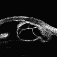 Idiopathic Iris Cyst
Aug 13 2025 by Virginia Gebhart
74 year old female with idiopathic iris cyst encroaching visual axis. Lesion is cystic with baseline internal reflectivity, unequivocal growth since first exam in 2022. Pt remains asymptomatic, will continue to observe.
Photographer: Virginia Gebhart, Retina Consultants of Carolina
Imaging device: Ellex Eye Cubed
Condition/keywords: cyst, cystic lesion, idiopathic cysts, ultrasound biomicroscopy
-
 Acute Retinal Necrosis (resolved)
Aug 21 2025 by Virginia Gebhart
31 year old female with resolved Acute Retinal Necrosis. Pt was positive for HSV2 in 2019, received intravitreal Ganciclovir x3. Both eyes remain stable with no active inflammation, retinitis remains inactive with pigmented edges. Pt continues taking Valtrex qday for maintenance. BCVA 20/40
Photographer: Virginia Gebhart, Retina Consultants of Carolina
Imaging device: Optos California
Condition/keywords: acute retinal necrosis, retinitis
-
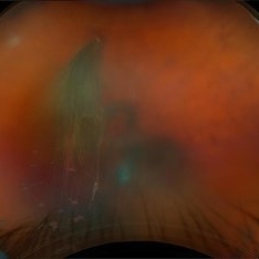 Retinal Tear w/VH
Aug 22 2025 by Virginia Gebhart
69 year old male referred for sudden vision loss. Difficult view secondary to VH. Ultrasound and photos show small break with clotting, heavy amount of layered blood inferior and scattered IRH's. Cryotherapy performed to seal current tear, will give VH time to clear on its own. Pt takes Eliquis twice a day.
Photographer: Virginia Gebhart, Retina Consultants of Carolina
Imaging device: Optos California
Condition/keywords: cryo-retinal tear, horseshoe tear, tear, vitreous hemorrhage
-
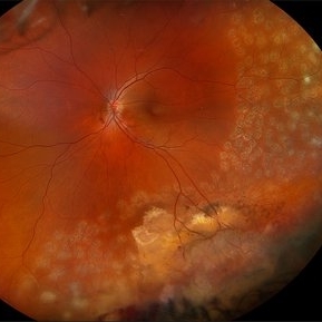 Vasoproliferative Tumor (FEVR) s/p PPV/PRP
Aug 27 2025 by Virginia Gebhart
39 year old female with an amelanotic vascular lesion inferotemporal with CR atrophy inferior edge and likely lipid exudate superior edge. Pt presented with vitreous and sub-hyaloid hemorrhage. Findings from exam, ultrasound, FA all consistent with FEVR, stage 2. PPV with PRP performed, pt vison has improved from CF@2ft at initial visit to 20/100 PH 20/60 at 1 week post-op. Pt's 2 children have been recently examined with identical findings of FEVR
Photographer: Virginia Gebhart, Retina Consultants of Carolina
Imaging device: Optos California
Condition/keywords: familial exudative vitreoretinopathy (FEVR), pan-retinal photocoagulation (PRP), Vasoproliferative Tumor
-
 Recurrent Ciliary Body Melanoma After 19 Years
Sep 10 2025 by Virginia Gebhart
86 year old female with recurrence of ciliary body melanoma s/p sectoral iridectomy in 2006. Small pigmented lesion has shown unequivocal growth since initial presentation in 2024. Unclear if this is a new lesion or has been present for an extended time. Based on exam, diagnostics, and review of records, the determination was made for treatment by placement of radiation plaque for 4 days.
Photographer: Virginia Gebhart, Retina Consultants of Carolina
Imaging device: Ellex Eye Cubed
Condition/keywords: ciliary body melanoma, melanoma, recurrence, ultrasound biomicroscopy
-
 Closed Funnel RD
Sep 26 2025 by Virginia Gebhart
B scan ultrasound of 12 year old male with complete closed funnel RD. Pt endorses several prior blunt traumas which may have caused detachment. If not secondary to trauma, may represent end-stage Coat's disease. FEVR unlikely due to FA findings of fellow eye. Eye is stable since initial visit in 2024, surgical intervention not recommended at this time. Vision NLP
Photographer: Virginia Gebhart, Retina Consultants of Carolina
Imaging device: Keeler Accutome
Condition/keywords: B scan ultrasound, chronic retinal detachment, Closed funnel RD, retinal detachment
-
 Choroidal Melanoma
Oct 1 2025 by Virginia Gebhart
60 year old male referred by optometrist for retinal detachment. Pt had been having symptoms of flashing lights and shadow in vision for approximately 1 month. Exam and diagnostics consistent with choroidal melanoma with exudative detachment inferior. Due to size of lesion (10.8 x 14.8 x 13.2) enucleation was recommended. Pt will be scheduled for surgery pending CT scan results.
Photographer: Virginia Gebhart, Retina Consultants of Carolina
Imaging device: Optos California
Condition/keywords: choroidal melanoma, exudative detachment, melanoma
-
 Choroidal Melanoma
Oct 3 2025 by Virginia Gebhart
63 year old male with new choroidal melanoma. Pt states vision has been poor for 2 years, worsening in the last year. Bscan ultrasound shows lesion extends into the macula up to the optic nerve. Recommended enucleation due to size of lesion (7.7 x 15.6 x 15.2) and poor prognosis of visual recovery. Surgery will be scheduled pending CT scan results.
Photographer: Virginia Gebhart, Retina Consultants of Carolina
Imaging device: Optos California
Condition/keywords: choroidal melanoma, exudative detachment
-
 Asteroid Hyalosis
Oct 8 2025 by Virginia Gebhart
49 year old female with asteroid hyalosis. Pt only notices when in bright light.
Photographer: Virginia Gebhart, Retina Consultants of Carolina
Imaging device: Optos California
Condition/keywords: asteroid hyalosis
-
Venous Pulsations in Large Choroidal Melanoma
Oct 9 2025 by Virginia Gebhart
81 year old female diagnosed with large, pigmented collar button tumor. Given size of lesion enucleation was recommended. CT scans show no evidence of metastatic disease. Pt is doing well s/p enucleation.
Condition/keywords: bscan ultrasound, choroidal melanoma
-
 Severe Papilledema
Oct 13 2025 by Virginia Gebhart
26 year old female with severe optic nerve edema OU with nerve fiber layer hemorrhages. Pt reports blurry vision, intermittent vision loss, and ongoing headaches. VF testing showed enlarged blind spots and possible early acuate defects OU. Pt sent to ER for MRI of brain to rule out tumor. Results pending.
Photographer: Virginia Gebhart, Retina Consultants of Carolina
Imaging device: Topcon TRC 50DX
Condition/keywords: idiopathic intracranial hypertension, optic nerve edema, papilledema
-
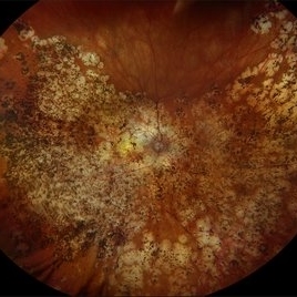 Multifocal Choroiditis with Panuveitis
Oct 16 2025 by Virginia Gebhart
39 year old female diagnosed with MCP in 2009. Extensive RPE changes and hypertrophy, arterial attenuation and pale nerve. Currently no active inflammation.
Photographer: Virginia Gebhart, Retina Consultants of Carolina
Imaging device: Optos California
Condition/keywords: corticosteroid-induced glaucoma, hypertrophy, multifocal chorioretinitis (MCP), PALE DISC
-
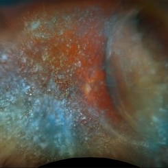 Choroidal and Near Total RD, Severe Asteroid Hyalosis, Treated Melanoma
Oct 22 2025 by Virginia Gebhart
78 year old male with sudden decrease in vision. Poor view due significant asteroid hyalosis. Bscan showed large nasal choroidal and near total retinal detachments, attached temporally. No obvious break found. Regressed tumor inferiorly s/p brachytherapy in April 2023. BCVA 20/320, IOP of 03. Pt schedule for primary PPV and possible SB placement vs. GFE
Photographer: Virginia Gebhart, Retina Consultants of Carolina
Imaging device: Optos California
Condition/keywords: asteroid hyalosis, brachytherapy, choroidal detachment, choroidal melanoma, melanoma, RD, retinal detachment, sub-total retinal detachment
-
 Vasoproliferative Tumor
Oct 27 2025 by Virginia Gebhart
34 year old male with retinal vasoproliferative tumor with traction detachment temporally and striae in the macula. Recommend vitrectomy prior to cryotherapy to release vitreous from retina. BCVA 20/30
Photographer: Virginia Gebhart, Retina Consultants of Carolina
Imaging device: Optos California
Condition/keywords: traction retinoschisis, tractional retinal detachment, Vasoproliferative Tumor
-
 Giant Nevus of the Macula
Nov 5 2025 by Virginia Gebhart
68 year old female with stable large pigmented lesion throughout the whole posterior pole, extending beyond the superior and inferior arcades. Pt has been aware of nevus for 45 years. Lesion has been stable since initial photos in 2013. Will continue to observe. Vision CF
Photographer: Virginia Gebhart, Retina Consultants of Carolina
Imaging device: Optos California
Condition/keywords: Choroidal nevus, pigmented lesion, pigmented nevus
-
 Bullous Retinal Detachment
Nov 13 2025 by Virginia Gebhart
42 year old female referred for vision loss x 4-5 days. Bullous retinal detachment from 8:00 to 3:00 with retinal tear at 11:00. Macula is detached. Vision is LP, IOP of 3. Pt is scheduled for GFE and possible scleral buckle.
Photographer: Virginia Gebhart, Retina Consultants of Carolina
Imaging device: Optos California
Condition/keywords: bullous retinal detachment, retinal detachment, retinal detachment of the macula, retinal tear with detachment
-
 Traction Detachment of Retina
Nov 14 2025 by Virginia Gebhart
50 year old female with proliferative diabetic retinopathy, large ridge of traction temporally, and significant band of fibrosis. Subretinal fluid throughout the macula. Due to traction, surgical repair not recommended at this time as it could worsen condition. Will observe closely. BCVA CF @ 1 ft.
Photographer: Virginia Gebhart, Retina Consultants of Carolina
Imaging device: Optos California
Condition/keywords: fibrosis, proliferative diabetic retinopathy (PDR), traction detachment, Traction retinal detachment
-
 PCO with Significant Capsular Distention
Nov 24 2025 by Virginia Gebhart
62 year old male presented with blurry vision x 3 weeks. UBM showed sulcus IOL with capsular distention, and possible aqueous misdirection into the capsule. Lens is being pushed into the iris. Pt referred back to cataract surgeon for consideration of YAG, may require PPV.
Photographer: Virginia Gebhart, Retina Consultants of Carolina
Imaging device: Ellex Eyecubed
Condition/keywords: aqueous misdirection, posterior capsule opacification
-
 CHRPE
CHRPE
Dec 11 2025 by Virginia Gebhart
48 year old female referred for pigmented lesion. Exam and photos consistent with well circumscribed CHRPE with lacunae. Patient previously unaware. Observation recommended.
Photographer: Virginia Gebhart, Retina Consultants of Carolina
Imaging device: Optos California
Condition/keywords: CHRPE, congenital hypertrophy of the retinal pigment epithelium (CHRPE)
-
 Nevus to Melanoma in 1 Year
Dec 12 2025 by Virginia Gebhart
83 year old male presents with partially amelanotic choroidal nevus, which has been followed for the last 10 years. On exam there is a new area of orange pigment, and change in diameter compared to photos from 2024. Ultrasound shows small increase in height, medium to low internal reflectivity, and vascularity. Pt is asymptomatic. Due to personal matters pt wishes to reassess in few months and schedule treatment.
Photographer: Virginia Gebhart, Retina Consultants of Carolina
Imaging device: Optos California
Condition/keywords: choroidal melanoma, choroidal nevus
-
 Peripheral Exudative Hemorrhagic Chorioretinopathy
Dec 17 2025 by Virginia Gebhart
77 year old female with amelanotic choroidal lesion with subretinal hemorrhage at the base and exudate inferiorly. Lesion has decreased on exam, photos and ultrasound after 4 months. Ultrasound showed irregular lesion that is regressing, variable internal reflectivity, and no venous pulsations. Findings most consistent with PEHCR. Will continue to observe.
Photographer: Virginia Gebhart, Retina Consultants of Carolina
Imaging device: Optos California
Condition/keywords: asteroid hyalosis, peripheral exudative hemorrhagic chorioretinopathy (PEHCR)
-
 New Collar Button Melanoma
Jan 7 2026 by Virginia Gebhart
67 year old female referred for possible choroidal melanoma. Clinical exam, photos and ultrasound findings consistent with melanoma. Large pigmented tumor with collar button configuration, subretinal fluid inferior and vitreous hemorrhage. Left eye has scleral pigmentation 360 degrees, pt states she has had "brown spots" on her eye since childhood. Iris and fundus are also noticeably darker in the left eye consistent with oculodermal melanocytosis. Pt states she has had regular eye exams and has never been diagnosed. Pt will be scheduled for plaque brachytherapy pending CT scan results.
Photographer: Virginia Gebhart, Retina Consultants of Carolina
Imaging device: Optos California
Condition/keywords: choroidal melanoma, collar button, hemorrhage, melanoma, Oculodermal Melanocytosis
-
 Ciliochoroidal Melanoma
Jan 15 2026 by Virginia Gebhart
54 year old female referred for choroidal lesion with possible retinal detachment. Pt states she did not notice any vision changes until she failed exam at the DMV. On exam there is a large pigmented ciliary body mass with a height measuring 13.2 mm. Ultrasound shows large mass with low internal reflectivity and vascularity. Due to tumor size enucleation was recommended. Pt will be scheduled for surgery pending CT scan results. Also ordered blood test HLA-A0201 to check eligibility for Kimmtrak if cancer spreads.
Photographer: Virginia Gebhart, Retina Consultants of Carolina
Imaging device: Optos California
Condition/keywords: ciliary body mass, ciliary body melanoma, ciliochoroidal melanoma
A project from the American Society of Retina Specialists