-
 Toxocara Granuloma
Jun 4 2014 by Henry J. Kaplan, MD
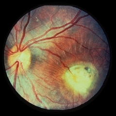
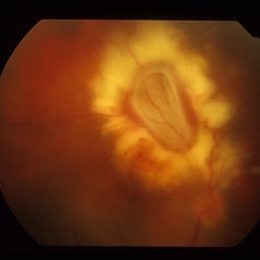
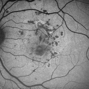
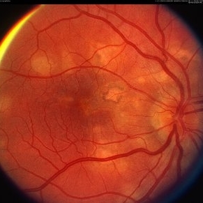
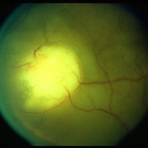
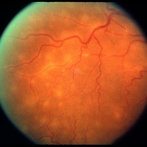
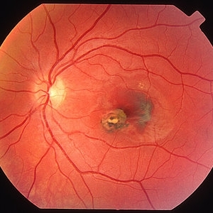
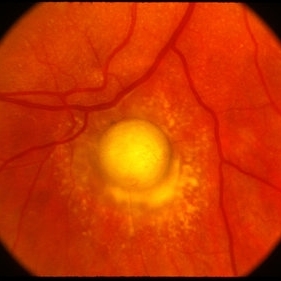
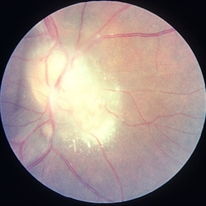
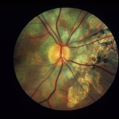
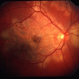
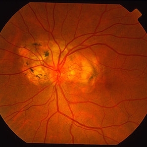
Toxocara granuloma in the macula OS. #1
Condition/keywords: toxocara granuloma
-
 ToxocaraGgranuloma
Jun 30 2014 by Henry J. Kaplan, MD
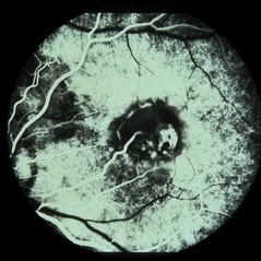
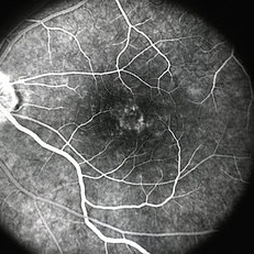
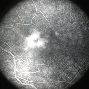
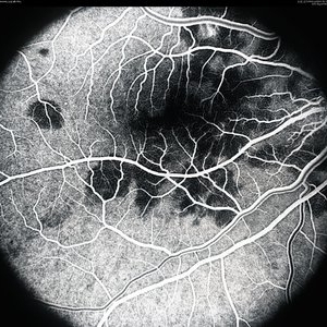
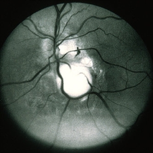
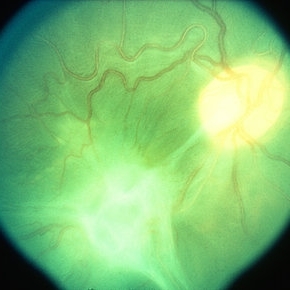
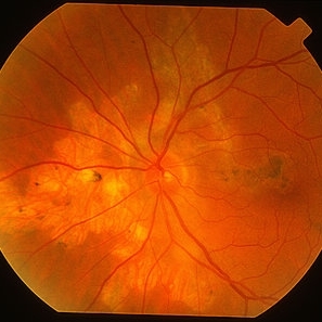
Arterial phase fluorescein angiography of the patient with toxocara granuloma shows hypofluorescence in most part of the lesion and arterial supply to the lesion #2.
Condition/keywords: toxocara granuloma
-
 Toxocara Granuloma
Jun 4 2014 by Henry J. Kaplan, MD
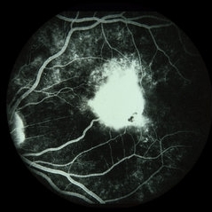
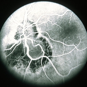
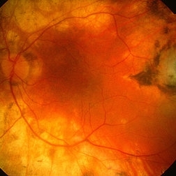
Arteriovenous phase angiogram of the same patient shows staining of the granuloma and stippling hyperfluorescence around the lesion secondary to RPE window defect. #3
Condition/keywords: toxocara granuloma, toxocariasis
-
 AIDS - Toxoplasmosis
Jun 4 2014 by Henry J. Kaplan, MD
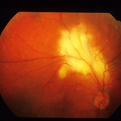
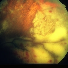
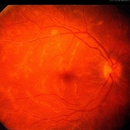
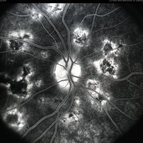
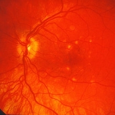
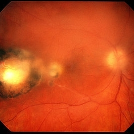
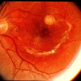
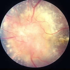
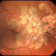
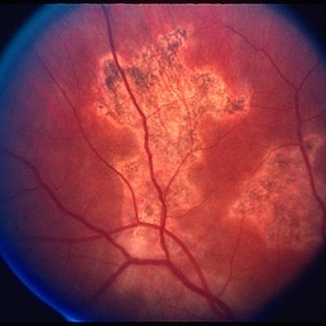
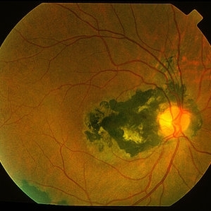
Fundus photograph of a patient with AIDS who has developed toxoplasma retinochoroiditis; large yellow patch of retinitis along the superior arcade fading the vasculature with feathery edges. #1
Condition/keywords: AIDS, toxoplasmosis
-
 AIDS - Toxoplasmosis
Jun 4 2014 by Henry J. Kaplan, MD
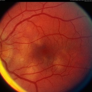
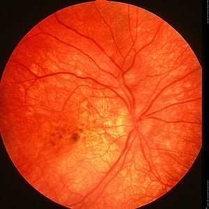
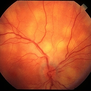
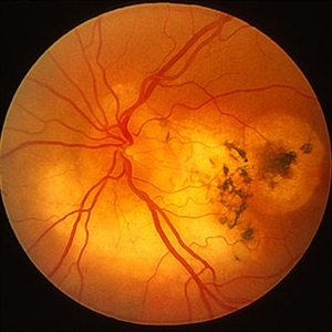
Fundus photograph of the same patient after treatment shows slow regression of the lesion. #2
Condition/keywords: AIDS, toxoplasmosis
-
 HLA-B27 Associated Uveitis
Jun 4 2014 by Henry J. Kaplan, MD
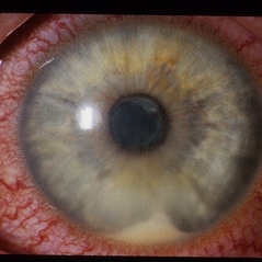
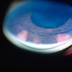
Severe anterior uveitis with fibrinous reaction and hypopyon formation related to HLA-B27. Notice the membrane on the lens surface.
Condition/keywords: acute anterior uveitis, HLA-B27, hypopyon
-
 Punctate Inner Choroidopathy Complicated with CNV
Jun 5 2013 by Henry J. Kaplan, MD
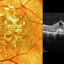
OCT of the same patient 6 weeks after Avastin injection shows decrease in intraretinal fluid and vision improved to 20/160. #4
Photographer: Angela Andersson
Imaging device: HRA II
Condition/keywords: choroidal neovascularization (CNV), punctate inner choroidopathy (PIC)
-
 Punctate Inner Choroidopathy Complicated with CNV
Jun 5 2013 by Henry J. Kaplan, MD
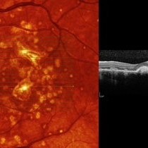
OCT of the same patient demonstrates CNV complex with intraretinal cystoid edema #3.
Photographer: Angela Andersson
Imaging device: HRA II
Condition/keywords: choroidal neovascularization (CNV), punctate inner choroidopathy (PIC)
-
 Punctate Inner Choroidopathy Complicated with CNV
Jun 5 2013 by Henry J. Kaplan, MD
Fundus autofluorescence of the same patient demonstrates multiple hypoautofluorescent spots compatible with the clinical lesions #2.
Photographer: Angela Andersson
Imaging device: HRA II
Condition/keywords: choroidal neovascularization (CNV), punctate inner choroidopathy (PIC)
-
---thumb.jpg/image-square;max$300,300.ImageHandler) Punctate Inner Choroidopathy Complicated with CNV
Punctate Inner Choroidopathy Complicated with CNV
Jun 5 2013 by Henry J. Kaplan, MD
A 15-year-old girl presented with blurred vision in left eye (20/200), fundus photography shows multiple deep round hypopigmented scars in the posterior pole with subretinal neovascular membrane #1.
Photographer: Angela Andersson
Condition/keywords: choroidal neovascularization (CNV), punctate inner choroidopathy (PIC)
-
 Wegener's Granulomatosis
May 2 2013 by Henry J. Kaplan, MD
Extensive peripheral retinitis and hemorrhagic foci in a patient with Wegner's granulomatosis.
Condition/keywords: Wegener's granulomatosis
-
 CME
CME
Mar 29 2013 by Henry J. Kaplan, MD
Typical cystic lesions in the fovea of a patient with CME.
Condition/keywords: cystoid macular edema (CME)
-
 CME
CME
Mar 29 2013 by Henry J. Kaplan, MD
Fluorescein angiogram of CME #1.
Condition/keywords: cystoid macular edema (CME)
-
 CME
CME
Mar 29 2013 by Henry J. Kaplan, MD
Fluorescein angiogram of CME, late phase, typical leakage as flower petaloid pattern #2.
Condition/keywords: cystoid macular edema (CME)
-
 Frosted Branch Angiitis
Mar 4 2013 by Henry J. Kaplan, MD
Frosted branch angiitis. Right Eye. #1
Condition/keywords: frosted branch angiitis
-
---thumb.jpg/image-square;max$300,300.ImageHandler) Frosted Branch Angiitis
Frosted Branch Angiitis
Feb 26 2013 by Henry J. Kaplan, MD
Frosted branch angiitis, left eye. #2
Condition/keywords: frosted branch angiitis
-
---thumb.jpg/image-square;max$300,300.ImageHandler) Frosted Branch Angiitis
Frosted Branch Angiitis
Feb 26 2013 by Henry J. Kaplan, MD
Frosted branch angiitis: left eye.
Condition/keywords: frosted branch angiitis
-
---thumb.jpg/image-square;max$300,300.ImageHandler) Frosted Branch Angiitis
Frosted Branch Angiitis
Feb 26 2013 by Henry J. Kaplan, MD
Frosted branch angiitis. F/A #1
Condition/keywords: frosted branch angiitis
-
---thumb.jpg/image-square;max$300,300.ImageHandler) Frosted Branch Angiitis
Frosted Branch Angiitis
Feb 26 2013 by Henry J. Kaplan, MD
Frosted branch angiitis patient. Left eye: F/A leakage seen as hyperfluorescence along involved vessels. #2
Condition/keywords: frosted branch angiitis
-
---thumb.jpg/image-square;max$300,300.ImageHandler) Frosted Branch Angiitis
Frosted Branch Angiitis
Feb 26 2013 by Henry J. Kaplan, MD
Frosted branch angiitis . F/A severe leakage along the involved vessles at late stage. #4
Condition/keywords: frosted branch angiitis
-
---thumb.jpg/image-square;max$300,300.ImageHandler) Frosted Branch Angiitis
Frosted Branch Angiitis
Feb 26 2013 by Henry J. Kaplan, MD
Frosted branch angiitis . F/A , very late leakage. #4
Condition/keywords: frosted branch angiitis
-
---thumb.jpg/image-square;max$300,300.ImageHandler) Acute Posterior Multifocal Placoid Pigment Epitheliopathy
Acute Posterior Multifocal Placoid Pigment Epitheliopathy
Feb 27 2013 by Henry J. Kaplan, MD
APMPPE fundus photographs. Right Eye multiple placoid yellowish subretinal lesions #1.
Condition/keywords: acute posterior multifocal placoid pigment epitheliopathy (APMPPE), white dot syndrome
-
---thumb.jpg/image-square;max$300,300.ImageHandler) Acute Posterior Multifocal Placoid Pigment Epitheliopathy
Acute Posterior Multifocal Placoid Pigment Epitheliopathy
Feb 27 2013 by Henry J. Kaplan, MD
APMPPE, fundus photographs. Left eye: Multiple placoid subretinal yellow - white lesions #2.
Condition/keywords: acute posterior multifocal placoid pigment epitheliopathy (APMPPE), white dot syndrome
-
---thumb.jpg/image-square;max$300,300.ImageHandler) Acute Posterior Placoid Pigment Epitheliopathy
Acute Posterior Placoid Pigment Epitheliopathy
Feb 27 2013 by Henry J. Kaplan, MD
APMPPE, milder form, posterior placoid yellowish lesions visible.
Condition/keywords: acute posterior multifocal placoid pigment epitheliopathy (APMPPE)
-
 Acute Posterior Placoid Pigment Epitheliopathy
Mar 4 2013 by Henry J. Kaplan, MD
APMPPE; right eye; transition from acute stage to residual scar formation in some of the lesions. #1
Condition/keywords: acute posterior multifocal placoid pigment epitheliopathy (APMPPE), white dot syndrome
-
 Acute Posterior Multifocal Placoid Pigment
Mar 4 2013 by Henry J. Kaplan, MD
Multiple posterior placoid subretinal lesions in the left eye #2.
Condition/keywords: acute posterior multifocal placoid pigment epitheliopathy (APMPPE), white dot syndrome
-
---thumb.jpg/image-square;max$300,300.ImageHandler) Acute Posterior Multifocal Placoid Pigment Epitheliopathy
Acute Posterior Multifocal Placoid Pigment Epitheliopathy
Feb 27 2013 by Henry J. Kaplan, MD
APMPPE, red free imaging: right eye #1.
Condition/keywords: acute posterior multifocal placoid pigment epitheliopathy (APMPPE), white dot syndrome
-
 APMPPE, Fluorescein Angiography
Feb 28 2013 by Henry J. Kaplan, MD
Early multiple hypofluorescent subretinal lesions in the early phases of F/A of a patient with APMPPE due to delayed choroidal filling and also masking effect of choroidal edema. #2
Condition/keywords: acute posterior multifocal placoid pigment epitheliopathy (APMPPE), white dot syndrome
-
---thumb.jpg/image-square;max$300,300.ImageHandler) Acute Posterior Multifocal Placoid Pigment Epitheliopathy
Acute Posterior Multifocal Placoid Pigment Epitheliopathy
Feb 27 2013 by Henry J. Kaplan, MD
APMPPE. F/A .Late hyperfluorescence and staining of the lesions apparent #3.
Condition/keywords: acute posterior multifocal placoid pigment epitheliopathy (APMPPE), white dot syndrome
-
---thumb.jpg/image-square;max$300,300.ImageHandler) Acute Posterior Multifocal Placoid Pigment Epitheliopathy Late Stage Scar Formation
Acute Posterior Multifocal Placoid Pigment Epitheliopathy Late Stage Scar Formation
Feb 27 2013 by Henry J. Kaplan, MD
APMPPE late stage scar formation. Right Eye Multiple scar formations occurs in some of the patients #1
Condition/keywords: acute posterior multifocal placoid pigment epitheliopathy (APMPPE), late stage, white dot syndrome
-
---thumb.jpg/image-square;max$300,300.ImageHandler) APMPPE Late Stage Scar Formation
APMPPE Late Stage Scar Formation
Feb 27 2013 by Henry J. Kaplan, MD
APMPPE late stage, multiple scar formation, left eye #2.
Condition/keywords: acute posterior multifocal placoid pigment epitheliopathy (APMPPE), late stage, white dot syndrome
-
---thumb.jpg/image-square;max$300,300.ImageHandler) Acute Posterior Multifocal Placoid Pigment Late Stage
Acute Posterior Multifocal Placoid Pigment Late Stage
Feb 27 2013 by Henry J. Kaplan, MD
APMPPE late stage scar formation.
Condition/keywords: acute posterior multifocal placoid pigment epitheliopathy (APMPPE), late stage, white dot syndrome
-
---thumb.jpg/image-square;max$300,300.ImageHandler) APMPPE Late Stage Scar Formation
APMPPE Late Stage Scar Formation
Feb 27 2013 by Henry J. Kaplan, MD
APMPPE late stage scar formation. F/A hypofluorescence in the lesions area is due to masking effect of pigments . #1
Condition/keywords: acute posterior multifocal placoid pigment epitheliopathy (APMPPE), late stage, white dot syndrome
-
 APMPPE, Late Stage, Scar Formation
Mar 4 2013 by Henry J. Kaplan, MD
APMPPE, late stage, scar formation, F/A #2
Condition/keywords: acute posterior multifocal placoid pigment epitheliopathy (APMPPE), white dot syndrome
-
---thumb.jpg/image-square;max$300,300.ImageHandler) APMPPE Late Stage Scar Formation
APMPPE Late Stage Scar Formation
Feb 27 2013 by Henry J. Kaplan, MD
APMPPE late stage scar formation. #3
Condition/keywords: acute posterior multifocal placoid pigment epitheliopathy (APMPPE), late stage, white dot syndrome
-
---thumb.jpg/image-square;max$300,300.ImageHandler) Roth Spot
Roth Spot
Feb 27 2013 by Henry J. Kaplan, MD
Roth spots due to subacute bacterial endocardiris in a patient with the diagnosis of AIDS .
Condition/keywords: AIDS, subacute bacterial endocardiris, white centered retinal hemorrhage (Roth Spot)
-
---thumb.jpg/image-square;max$300,300.ImageHandler) Roth Spot
Roth Spot
Feb 27 2013 by Henry J. Kaplan, MD
Roth spot due to subacute bacterial endocardiris in AIDS patient. Magnified view in the same patient.
Condition/keywords: AIDS, subacute bacterial endocardiris, white centered retinal hemorrhage (Roth Spot)
-
 Ophthalmomyasis
May 2 2013 by Henry J. Kaplan, MD
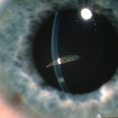
Floating larva in the anterior chamber.
Condition/keywords: ophthalmomyiasis
-
---thumb.jpg/image-square;max$300,300.ImageHandler) Ophthalmomyasis
Ophthalmomyasis
-
---thumb.jpg/image-square;max$300,300.ImageHandler) CMV Retinitis in AIDS
CMV Retinitis in AIDS
Feb 27 2013 by Henry J. Kaplan, MD
CMV retinitis in a patient with the diagnosis of AIDS: relatively early stage; at the beginning the lesions may look like a cotton wool spot ; one of them is seen superotemporal to the optic disc.
Condition/keywords: AIDS
-
---thumb.jpg/image-square;max$300,300.ImageHandler) CMV Retinitis in a Patient with the Diagnosis of AIDS
CMV Retinitis in a Patient with the Diagnosis of AIDS
Feb 27 2013 by Henry J. Kaplan, MD
Color fundus photograph, right eye: CMV neuroretinitis (retinitis which begins from the arcades and is accompanied by hemorrhage and also has involved the optic nerve).
Condition/keywords: AIDS
-
---thumb.jpg/image-square;max$300,300.ImageHandler) CMV Retinitis in a Patient with the Diagnosis of AIDS
CMV Retinitis in a Patient with the Diagnosis of AIDS
Feb 27 2013 by Henry J. Kaplan, MD
CMV retinitis, left eye: classic form in AIDS patient. Hemorrhagic retinitis mainly in the superior arcade.
Condition/keywords: AIDS
-
---thumb.jpg/image-square;max$300,300.ImageHandler) CMV Frosted Branch Angitis
CMV Frosted Branch Angitis
Feb 27 2013 by Henry J. Kaplan, MD
Fundus photograph: development of frosted branch angitis in the retina of a patient with CMV retinitis.
Condition/keywords: frosted branch angiitis
-
---thumb.jpg/image-square;max$300,300.ImageHandler) CMV Retinitis Granular Type
CMV Retinitis Granular Type
Feb 27 2013 by Henry J. Kaplan, MD
CMV retinitis; granular pigmentary changes are seen along with frosted branch angitis.
Condition/keywords: frosted branch angiitis
-
---thumb.jpg/image-square;max$300,300.ImageHandler) CMV Inclusion Bodies
CMV Inclusion Bodies
Feb 27 2013 by Henry J. Kaplan, MD
Owl's eye inclusion bodies in CMV retinitis
-
---thumb.jpg/image-square;max$300,300.ImageHandler) CMV Retinitis
CMV Retinitis
Feb 27 2013 by Henry J. Kaplan, MD
Classic CMV retinitis in the left eye: before treatment. #1
-
---thumb.jpg/image-square;max$300,300.ImageHandler) CMV Retinitis
CMV Retinitis
Feb 27 2013 by Henry J. Kaplan, MD
CMV retinitis, gradual improvement after treatment, with intravenous ganciclovir #2.
Condition/keywords: ganciclovir
-
---thumb.jpg/image-square;max$300,300.ImageHandler) CMV Retinitis
CMV Retinitis
Feb 27 2013 by Henry J. Kaplan, MD
CMV retinitis, improvement after treatment with ganciclovir #3.
Condition/keywords: after treatment, ganciclovir
-
---thumb.jpg/image-square;max$300,300.ImageHandler) CMV Retinitis
CMV Retinitis
Feb 27 2013 by Henry J. Kaplan, MD
CMV retinitis, almost resolved after treatment with ganciclovir #4.
Condition/keywords: after treatment, ganciclovir
-
---thumb.jpg/image-square;max$300,300.ImageHandler) ARN
ARN
Feb 27 2013 by Henry J. Kaplan, MD
ARN; hazy media is due to moderate to severe vitritis
Condition/keywords: acute retinal necrosis
-
---thumb.jpg/image-square;max$300,300.ImageHandler) ARN
ARN
Feb 27 2013 by Henry J. Kaplan, MD
Peripheral necrotizing retinitis in an ARN patient.
Condition/keywords: acute retinal necrosis
-
---thumb.jpg/image-square;max$300,300.ImageHandler) ARN
ARN
Feb 27 2013 by Henry J. Kaplan, MD
ARN, severe vitritis, and necrotizing retina.
Condition/keywords: acute retinal necrosis, necrotizing retina, vitritis
-
---thumb.jpg/image-square;max$300,300.ImageHandler) ARN : Zoster Intranuclear Inclusion Bodies
ARN : Zoster Intranuclear Inclusion Bodies
Feb 27 2013 by Henry J. Kaplan, MD
ARN, zoster intranuclear inclusion bodies.
Condition/keywords: acute retinal necrosis, inclusion bodies, varicella zoster virus (VZV)
-
---thumb.jpg/image-square;max$300,300.ImageHandler) ARN
ARN
Feb 27 2013 by Henry J. Kaplan, MD
ARN, VZV intranuclear inclusion bodies (cowdry bodies).
Condition/keywords: acute retinal necrosis
-
---thumb.jpg/image-square;max$300,300.ImageHandler) Complications of ARN
Complications of ARN
Feb 27 2013 by Henry J. Kaplan, MD
Complications of ARN. NVD in the right eye of a patient with a history of ARN.
Condition/keywords: acute retinal necrosis, neovascularization of the disc (NVD)
-
---thumb.jpg/image-square;max$300,300.ImageHandler) Complications of ARN
Complications of ARN
Feb 27 2013 by Henry J. Kaplan, MD
Complications of ARN: NVD as a late complication in the right eye of a patient with previous ARN.
Condition/keywords: ARN complications, neovascularization of the disc (NVD)
-
---thumb.jpg/image-square;max$300,300.ImageHandler) Complications of ARN, TRD
Complications of ARN, TRD
Feb 27 2013 by Henry J. Kaplan, MD
Development of TRD as a late complication of ARN.
Condition/keywords: acute retinal necrosis, ARN complications, tractional retinal detachment
-
---thumb.jpg/image-square;max$300,300.ImageHandler) HIV Retinopathy
HIV Retinopathy
Feb 27 2013 by Henry J. Kaplan, MD
HIV retinopathy, multiple cotton wool spots, right eye. #1
Condition/keywords: cotton wool spots, HIV retinopathy
-
---thumb.jpg/image-square;max$300,300.ImageHandler) HIV Retinopathy
HIV Retinopathy
Feb 27 2013 by Henry J. Kaplan, MD
HIV retinopathy, left eye: multiple cotton wool spots. #2
Condition/keywords: cotton wool spots, HIV retinopathy
-
---thumb.jpg/image-square;max$300,300.ImageHandler) Candida Endophthalmitis
Candida Endophthalmitis
Feb 26 2013 by Henry J. Kaplan, MD
Candida endophthalmitis, (typical string of pearls in the vitreous).
Condition/keywords: candida endophthalmitis, string of pearls
-
---thumb.jpg/image-square;max$300,300.ImageHandler) Presumed Ocular Histoplasmosis Syndrome
Presumed Ocular Histoplasmosis Syndrome
Feb 26 2013 by Henry J. Kaplan, MD
Color fundus photograph of the right eye of a patient with POHS shows typical punched out scars and peripapillary atrophy.
Condition/keywords: presumed ocular histoplasmosis syndrome (POHS)
-
---thumb.jpg/image-square;max$300,300.ImageHandler) POHS Complicated by CNV
POHS Complicated by CNV
Feb 26 2013 by Henry J. Kaplan, MD
POHS complicated by CNV: subretinal hemorrhage in the fovea adjacent to POHS scar.
Condition/keywords: choroidal neovascularization (CNV), presumed ocular histoplasmosis syndrome (POHS), subretinal hemorrhage
-
---thumb.jpg/image-square;max$300,300.ImageHandler) Presumed Ocular Histoplasmosis Syndrome
Presumed Ocular Histoplasmosis Syndrome
Feb 26 2013 by Henry J. Kaplan, MD
POHS; left eye: large punched out pigmented scars and peripapillary atrophy.
Condition/keywords: presumed ocular histoplasmosis syndrome (POHS)
-
---thumb.jpg/image-square;max$300,300.ImageHandler) Old Presumed Ocular Histoplasmosis Syndrome
Old Presumed Ocular Histoplasmosis Syndrome
Feb 26 2013 by Henry J. Kaplan, MD
Old POHS, advanced subretinal scar formation due to CNV (end stage).
Condition/keywords: end stage, presumed ocular histoplasmosis syndrome (POHS), subretinal scar formation
-
---thumb.jpg/image-square;max$300,300.ImageHandler) Presumed Ocular Histoplasmosis Syndrome
Presumed Ocular Histoplasmosis Syndrome
Feb 26 2013 by Henry J. Kaplan, MD
POHS complicated by CNV and subretinal hemorrhage in fovea.
Condition/keywords: choroidal neovascularization (CNV), presumed ocular histoplasmosis syndrome (POHS)
-
---thumb.jpg/image-square;max$300,300.ImageHandler) Presumed Ocular Histoplasmosis Syndrome
Presumed Ocular Histoplasmosis Syndrome
Feb 26 2013 by Henry J. Kaplan, MD
POHS complicated by CNV and subretinal hemorrhage in the extrafoveal area superiorly.
Condition/keywords: choroidal neovascularization (CNV), presumed ocular histoplasmosis syndrome (POHS), subretinal hemorrhage
-
---thumb.jpg/image-square;max$300,300.ImageHandler) Birdshot Retinochoroidopathy
Birdshot Retinochoroidopathy
Feb 27 2013 by Henry J. Kaplan, MD
Birdshot retinochoroidopathy; right Eye; #1.
Condition/keywords: birdshot retinochoroidopathy, white dot syndrome
-
---thumb.jpg/image-square;max$300,300.ImageHandler) Birdshot Retinochoroidopathy
Birdshot Retinochoroidopathy
Feb 26 2013 by Henry J. Kaplan, MD
Birdshot retinochoroidopathy; left eye: acute stage, haze due to vitritis and multiple oval cream colored lesions at the level of choroid.
Condition/keywords: birdshot retinochoroidopathy
-
---thumb.jpg/image-square;max$300,300.ImageHandler) Birdshot Retinochoroidopathy
Birdshot Retinochoroidopathy
Feb 26 2013 by Henry J. Kaplan, MD
Birdshot retinochoroidopathy.
Condition/keywords: birdshot retinochoroidopathy, white dot syndrome
-
---thumb.jpg/image-square;max$300,300.ImageHandler) Birdshot Retinochoroidopathy
Birdshot Retinochoroidopathy
Feb 26 2013 by Henry J. Kaplan, MD
Birdshot retinochoroidopathy: multiple cream colored oval lesions most prominant on the nasal retina.
Condition/keywords: birdshot, birdshot retinochoroidopathy
-
---thumb.jpg/image-square;max$300,300.ImageHandler) Birdshot Retinochoroidopathy
Birdshot Retinochoroidopathy
Feb 26 2013 by Henry J. Kaplan, MD
Birdshot retinochoroidopathy. Multiple ovoid yellow choroidal lesions spread out radially most prominent on nasal side.
Condition/keywords: birdshot, birdshot retinochoroidopathy
-
---thumb.jpg/image-square;max$300,300.ImageHandler) Birdshot Retinochoroidopathy
Birdshot Retinochoroidopathy
Feb 27 2013 by Henry J. Kaplan, MD
Birdshot retinochoroidopathy.
Condition/keywords: birdshot retinochoroidopathy
-
---thumb.jpg/image-square;max$300,300.ImageHandler) Vogt koyanagi Harrada Depigmentations
Vogt koyanagi Harrada Depigmentations
Feb 26 2013 by Henry J. Kaplan, MD
Vitiligo and poliosis in a patient with VKH syndrome.
-
---thumb.jpg/image-square;max$300,300.ImageHandler) Vogt Koyanagi Harrada Acute Stage
Vogt Koyanagi Harrada Acute Stage
Feb 26 2013 by Henry J. Kaplan, MD
VKH acute stage; bilateral multiple serous detachments in the same patient with vitiligo and poliosis.
-
---thumb.jpg/image-square;max$300,300.ImageHandler) Diffuse Unilateral Subacute Neuroretinitis
Diffuse Unilateral Subacute Neuroretinitis
Feb 26 2013 by Henry J. Kaplan, MD
DUSN typical track and large, subretinal.
Condition/keywords: diffuse unilateral subacute neuroretinitis (DUSN)
-
---thumb.jpg/image-square;max$300,300.ImageHandler) Diffuse Unilateral Subacute Neuroretinitis
Diffuse Unilateral Subacute Neuroretinitis
Feb 26 2013 by Henry J. Kaplan, MD
DUSN typical track and larva, subretinal.
Condition/keywords: diffuse unilateral subacute neuroretinitis (DUSN)
-
 Koeppe nodules
May 2 2013 by Henry J. Kaplan, MD
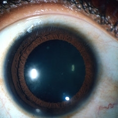
Granulomatous Koeppe nodules at the pupillary margin.
Condition/keywords: iris nodules, Koeppe nodules
-
---thumb.jpg/image-square;max$300,300.ImageHandler) Intermediate Uveitis
Intermediate Uveitis
Feb 26 2013 by Henry J. Kaplan, MD
Intermediate uveitis, virreous veils.
Condition/keywords: intermediate uveitis, virreous veils
-
 Berlin's Nodules
May 2 2013 by Henry J. Kaplan, MD
Granulomatous berlin's nodules in the angle secondary to sarcoidosis.
Condition/keywords: Berlin's nodules, sarcoidosis
-
---thumb.jpg/image-square;max$300,300.ImageHandler) Intermediate Uveitis
Intermediate Uveitis
Feb 26 2013 by Henry J. Kaplan, MD
Intermediate uveitis: shadow of vitreous veils visible on the retina; CME is also present.
Condition/keywords: cystoid macular edema (CME), intermediate uveitis, virreous veils
-
 Busacca nodules
May 2 2013 by Henry J. Kaplan, MD
Typical Busacca iris stromal nodules in sarcoid uveitis; notice the ps formation.
Condition/keywords: busacca nodulaes, granulomatous uveitis, iris nodules, sarcoid bussaca iris nodules
-
---thumb.jpg/image-square;max$300,300.ImageHandler) Intermediate Uveitis, Ciliary Body Cyst
Intermediate Uveitis, Ciliary Body Cyst
Feb 26 2013 by Henry J. Kaplan, MD
Intermediate uveitis, ciliary body cyst.
Condition/keywords: ciliary body cyst, intermediate uveitis
-
---thumb.jpg/image-square;max$300,300.ImageHandler) Intermediate Uveitis and CME
Intermediate Uveitis and CME
Feb 26 2013 by Henry J. Kaplan, MD
Cystoid macular edema in intermediate uveitis.
Condition/keywords: cystoid macular edema (CME), intermediate uveitis
-
 Punctate inner choroiditis
May 2 2013 by Henry J. Kaplan, MD
Multiple hypopigmented round spots in the posterior pole
Condition/keywords: punctate inner choroidopathy (PIC)
-
---thumb.jpg/image-square;max$300,300.ImageHandler) Intermediate Uveiris and CME
Intermediate Uveiris and CME
Feb 26 2013 by Henry J. Kaplan, MD
Fluorescein angiography of a patient with CME with typical flower petaloid leakage at late phase.
Condition/keywords: cystoid macular edema (CME), intermediate uveitis
-
---thumb.jpg/image-square;max$300,300.ImageHandler) SLE Vasculitis
SLE Vasculitis
Feb 26 2013 by Henry J. Kaplan, MD
SLE vasculitis complicated by CRAO; right eye. #1
Condition/keywords: central retinal artery occlusion (CRAO), systemic lupus erythematosus (SLE) retinopathy, systemic lupus erythematosus (SLE) vasculitis
-
---thumb.jpg/image-square;max$300,300.ImageHandler) SLE Vasculitis
SLE Vasculitis
Feb 26 2013 by Henry J. Kaplan, MD
SLE vasculitis complicated by CRAO; left eye of the same case. #2
Condition/keywords: central retinal artery occlusion (CRAO), systemic lupus erythematosus (SLE) vasculitis
-
---thumb.jpg/image-square;max$300,300.ImageHandler) Lupus Anticoagulant Disorder
Lupus Anticoagulant Disorder
Feb 26 2013 by Henry J. Kaplan, MD
Vasoocclusive retinopathy associated with Lupus anticoagulate factor.
Condition/keywords: lupus anticoagulate factor, vasoocclusive retinopathy
-
---thumb.jpg/image-square;max$300,300.ImageHandler) Lupus Anticoagulant Disorder
Lupus Anticoagulant Disorder
Feb 26 2013 by Henry J. Kaplan, MD
Occluded retinal vessels and vitreous hemorrhage apparent.
Condition/keywords: lupus anticoagulate factor
-
---thumb.jpg/image-square;max$300,300.ImageHandler) SLE Retinopathy
SLE Retinopathy
Feb 26 2013 by Henry J. Kaplan, MD
Cotton wool spots, right eye. #1
Condition/keywords: lupus, systemic lupus erythematosus (SLE) retinopathy, systemic lupus erythematosus (SLE) vasculitis
-
---thumb.jpg/image-square;max$300,300.ImageHandler) SLE Retinopathy
SLE Retinopathy
Feb 26 2013 by Henry J. Kaplan, MD
SLE retinopathy,l eft eye: multiple cotton wool spots and blot hemorrhages. #2
Condition/keywords: blot hemorrhages, cotton wool spots, systemic lupus erythematosus (SLE) retinopathy
-
 Vogt Koyanagi Harrada Late Stage
Mar 1 2013 by Henry J. Kaplan, MD
Right Eye: sun set glow appearance.
-
---thumb.jpg/image-square;max$300,300.ImageHandler) Vogt Koyanagi Harada Late Stage
Vogt Koyanagi Harada Late Stage
Feb 26 2013 by Henry J. Kaplan, MD
VKH late stage, left eye: sun set glow appearance. #2
-
---thumb.jpg/image-square;max$300,300.ImageHandler) Multifocal Choroiditis and Panuveitis Syndrome
Multifocal Choroiditis and Panuveitis Syndrome
Feb 26 2013 by Henry J. Kaplan, MD
Multifocal choroiditis and panuveitis: left eye. Acute stage: haziness of the media due to vitritis and multiple active yellow and also inactive choroidal lesions are present.
Condition/keywords: multifocal choroiditis
-
---thumb.jpg/image-square;max$300,300.ImageHandler) Multifocal Choroiditis & Panuveitis Syndrome
Multifocal Choroiditis & Panuveitis Syndrome
Feb 26 2013 by Henry J. Kaplan, MD
Multifocal choroiditis, MFC, old scars.
Condition/keywords: multifocal choroiditis, panuveitis
-
---thumb.jpg/image-square;max$300,300.ImageHandler) Multifocal Choroiditis and Panuveitis Syndrome
Multifocal Choroiditis and Panuveitis Syndrome
Feb 26 2013 by Henry J. Kaplan, MD
Multifocal choroiditis, left eye: multiple punched out scar formations in the posterior pole.
Condition/keywords: multifocal choroiditis, panuveitis
-
---thumb.jpg/image-square;max$300,300.ImageHandler) Multifocal Choroiditis & Panuveitis Syndrome
Multifocal Choroiditis & Panuveitis Syndrome
Feb 26 2013 by Henry J. Kaplan, MD
Multifocal choroiditis, MFC. Lesions look similar to POHS but the patient has vitritis in contrast to the former.
Condition/keywords: multifocal choroiditis, panuveitis
-
---thumb.jpg/image-square;max$300,300.ImageHandler) Multifocal Choroiditis
Multifocal Choroiditis
Feb 26 2013 by Henry J. Kaplan, MD
Multifocal choroiditis, MFC, inactive scars in the periphery.
Condition/keywords: multifocal choroiditis
-
---thumb.jpg/image-square;max$300,300.ImageHandler) Multifocal Choroiditis & Panuveitis Syndrome
Multifocal Choroiditis & Panuveitis Syndrome
Feb 26 2013 by Henry J. Kaplan, MD
Multifocal choroiditis (MFC) and panuveitis.
Condition/keywords: multifocal choroiditis, panuveitis
-
---thumb.jpg/image-square;max$300,300.ImageHandler) Lyme Disease
Lyme Disease
Feb 26 2013 by Henry J. Kaplan, MD
Lyme disease, tick on ear. #1 Skin and ophthalmic lesions in the following slides.
Condition/keywords: Lyme disease
-
---thumb.jpg/image-square;max$300,300.ImageHandler) Lyme Disease
Lyme Disease
Feb 26 2013 by Henry J. Kaplan, MD
Lyme disease, black-legged tick on ear. #2
Condition/keywords: Lyme disease
-
---thumb.jpg/image-square;max$300,300.ImageHandler) Lyme Disease
Lyme Disease
Feb 26 2013 by Henry J. Kaplan, MD
Lyme disease: spirochaete, or "corkscrew-shaped" Borrelia burgdorferi are visible by special staining during darkfield microscopy. #3
Condition/keywords: Lyme disease
-
---thumb.jpg/image-square;max$300,300.ImageHandler) Lyme Disease
Lyme Disease
-
---thumb.jpg/image-square;max$300,300.ImageHandler) Lyme Disease
Lyme Disease
Feb 26 2013 by Henry J. Kaplan, MD
Lyme disease, erythema migrans and typical annular lesions. #4
Condition/keywords: Lyme disease
-
---thumb.jpg/image-square;max$300,300.ImageHandler) Lyme Disease
Lyme Disease
Feb 26 2013 by Henry J. Kaplan, MD
Lyme disease, intermediate uveitis: severe vitritis and snow ball formations. #6
Condition/keywords: Lyme disease
-
 Tuberculosis Granuloma of the Choroid
Feb 25 2013 by Henry J. Kaplan, MD
Chroioidal tuberculoma.
Condition/keywords: choroidal tuberculoma, tuberculosis
-
 Tuberculosis Panuveitis
Feb 25 2013 by Henry J. Kaplan, MD
Peripheral vascular sheathing and multiple choroidiris foci in a patient with tuberculosis panuveitis.
Condition/keywords: panuveitis, tuberculosis
-
 OcularToxoplasmosis
Feb 25 2013 by Henry J. Kaplan, MD
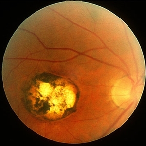
Toxoplasmosis, right eye: congenital typical macular scar with peripheral hyperpigmentation.
Condition/keywords: inactive toxoplasmosis, toxoplasmosis
-
 Toxoplasma Retinochoroiditis
Feb 25 2013 by Henry J. Kaplan, MD
Toxoplasmosis, right eye: reactivation of congenital toxoplasmosis as an active retinitis lesion with overlying vitritis adjacent to an old scar.
Condition/keywords: toxoplasmosis chorioretinitis, toxoplasmosis reactivation
-
 Toxoplasma Neuroretinitis (Jensen`s Disease)
Feb 25 2013 by Henry J. Kaplan, MD
Toxoplasma neuroretinitis in the left eye of a patient with macular star formation, retinitis adjacent to the optic nerve head with disc swelling.
Condition/keywords: Jensen disease, ocular toxoplasmosis, toxoplasmosis
-
 Secondary Choroidal Neovascularization Due to Toxoplasmosis
Feb 25 2013 by Henry J. Kaplan, MD
Left eye: secondary choroidal neovascularization and subretinal hemorrhage in a patient with old macular scar of toxoplasma.
Condition/keywords: choroidal neovascularization (CNV), toxoplasmosis, toxoplasmosis chorioretinitis
-
 Toxocara Granuloma
Feb 25 2013 by Henry J. Kaplan, MD
Toxocara granuloma of optic nerve head. Red free image #1; the rest of F/A in the following slides.
Condition/keywords: toxocara granuloma
-
 Toxocara Granuloma
Feb 25 2013 by Henry J. Kaplan, MD
Toxocara granuloma of optic nerve; F/A.
Condition/keywords: ocular toxoplasmosis, toxocara granuloma, toxocariasis
-
 Toxocara Granuloma
Feb 25 2013 by Henry J. Kaplan, MD
Toxocara granuloma of ON, late stage F/A. #3 Late hyperfluorescence in the granuloma due to staining.
Condition/keywords: ocular toxoplasmosis, toxocara granuloma, toxocariasis
-
 Toxocara Granuloma
Feb 25 2013 by Henry J. Kaplan, MD
Toxocara granuloma , epiretinal membrane and localized tractional detachment.
Condition/keywords: toxocara granuloma
-
 Toxocara Granuloma
Feb 25 2013 by Henry J. Kaplan, MD
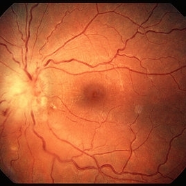
Toxocara granuloma superotemporal to the fovea.
Condition/keywords: ocular toxoplasmosis, toxocara granuloma, toxocariasis
-
 Toxocara Granuloma
Feb 25 2013 by Henry J. Kaplan, MD
Toxocara granuloma in the midperiphery of the retina.
Condition/keywords: ocular toxoplasmosis, toxocara granuloma, toxocariasis
-
 Toxocara Granuloma
Feb 25 2013 by Henry J. Kaplan, MD
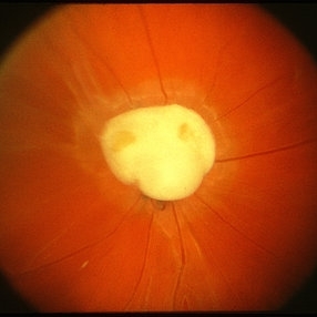
Toxocara granuloma of the optic nerve head.
Condition/keywords: ocular toxoplasmosis, toxocara granuloma, toxocariasis
-
 Bilateral Optic Nerve Involvement in Sarcoidosis
Feb 25 2013 by Henry J. Kaplan, MD
Optic nerve granuloma of sarcoidosis in the right eye of a patient with bilateral involvement #1. Left eye is in the following slide.
Condition/keywords: bilateral involvement, sarcoid granuloma
-
 Bilateral Optic Nerve Involvement in Sarcoidosis
Feb 25 2013 by Henry J. Kaplan, MD
Optic nerve head granuloma of sarcoidosis with severe infiltration and exudation in the left eye of the same patient #2.
Condition/keywords: bilateral involvement, sarcoid granuloma
-
 Sarcoidosis Choroiditis
Feb 25 2013 by Henry J. Kaplan, MD
Sarcoidosis multifocal choroiditis in a case with a known diagnosis of sarcoidosis.
Condition/keywords: sarcoidosis choroiditis
-
 Sarcoidosis
Feb 25 2013 by Henry J. Kaplan, MD
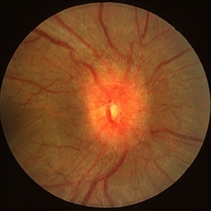
Optic nerve head infiltration of sarcoidosis presenting as optic nerve swelling.
Condition/keywords: sarcoidosis
-
 Serpiginous Choroiditis
Jun 4 2014 by Henry J. Kaplan, MD
typical serpentine lesions of serpiginous choroiditis. OD #1
Condition/keywords: serpiginous choroiditis
-
 Serpiginous Choroiditis
Jun 4 2014 by Henry J. Kaplan, MD
The same patients more nasally demonstrates scar formation and outer retinal atrophy. #2
Condition/keywords: serpiginous choroiditis
-
 Serpiginous Choroiditis
Feb 25 2013 by Henry J. Kaplan, MD
Typical serpiginous choroiditis: right eye.
Condition/keywords: serpiginous choroiditis
-
 Serpiginous Choroiditis
Feb 25 2013 by Henry J. Kaplan, MD
Serpiginous choroiditis, left eye. Active yellowish edematous lesion visible around the optic nerve toward the fovea and also old pigmented scar in the fovea.
Condition/keywords: serpiginous choroiditis
-
 Serpiginous Choroiditis
Feb 25 2013 by Henry J. Kaplan, MD
Serpiginous choroiditis. Typical feature going around the arcades.
Condition/keywords: serpiginous choroiditis
-
 Serpiginous Choroiditis
Feb 25 2013 by Henry J. Kaplan, MD
Serpiginous choroiditis, right eye. Both active and inactive lesions clearly visible; active lesions are the yellowish subretinal area most prominant nasal to optic nerve head and also around the inferior arcade and temporal to the macular lesion.
Condition/keywords: serpiginous choroiditis
-
 Serpiginous Choroiditis
Feb 25 2013 by Henry J. Kaplan, MD
Typical serpentine like lesion.
Condition/keywords: serpiginous choroiditis
-
 Serpiginous Choroiditis
Feb 25 2013 by Henry J. Kaplan, MD
Serpiginous choroiditis. (macula spared)
Condition/keywords: serpiginous choroiditis
-
 Serpiginous Choroiditis
Feb 25 2013 by Henry J. Kaplan, MD
Serpiginous choroiditis, right eye #1. Advanced pigmented scar formation ( fovea involved).
Condition/keywords: serpiginous choroiditis
-
 Serpiginous Choroiditis
Feb 25 2013 by Henry J. Kaplan, MD
Serpiginous choroiditis, left eye #2. Active edematous lesion visible moving toward the fovea.
Condition/keywords: serpiginous choroiditis
A project from the American Society of Retina Specialists