-
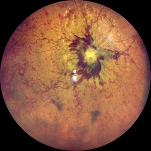
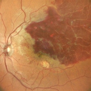
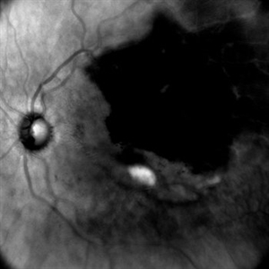
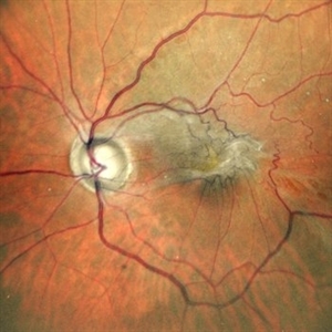
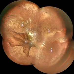
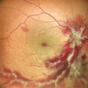
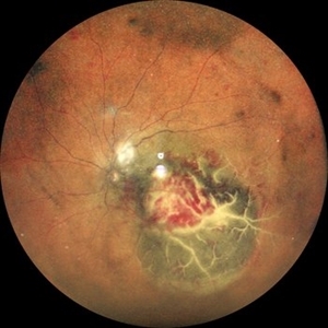
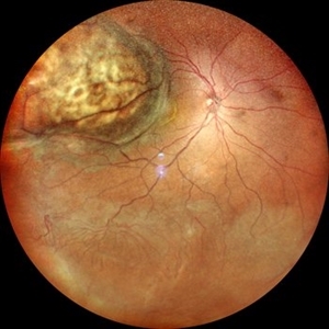
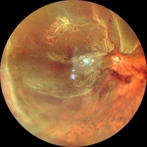
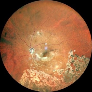
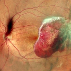
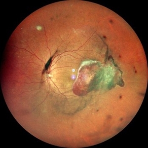
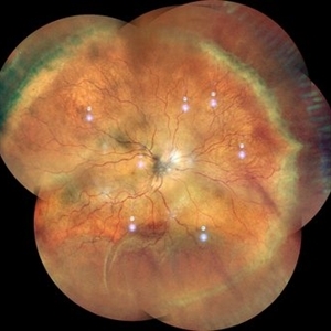
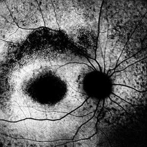
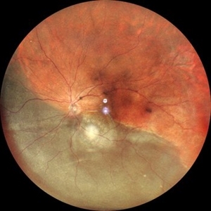
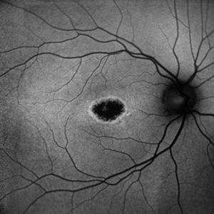
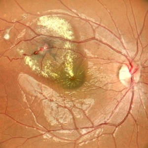
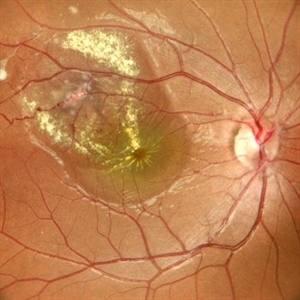
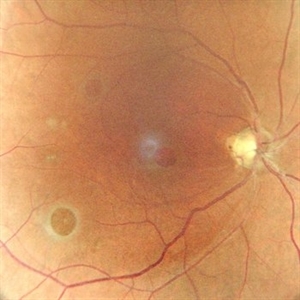
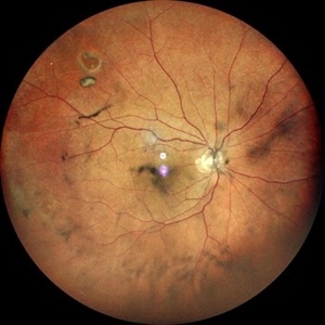
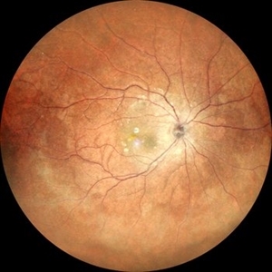
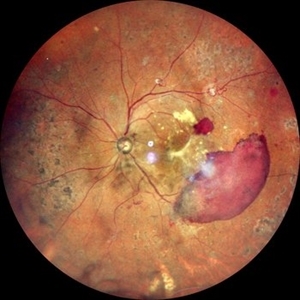
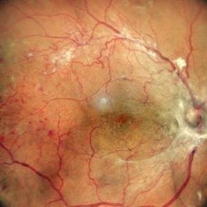
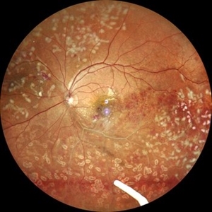
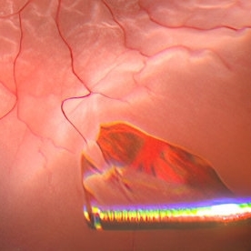
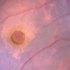
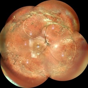
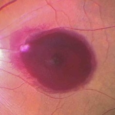
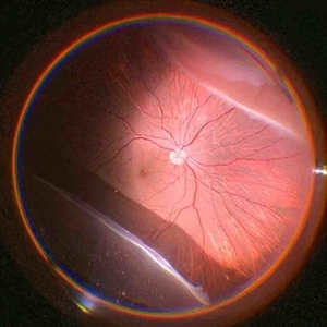
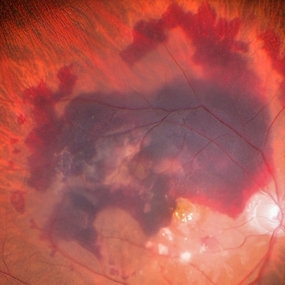
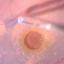
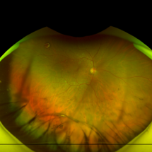
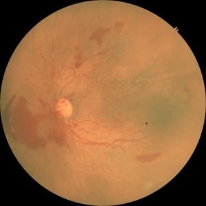
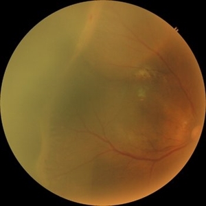
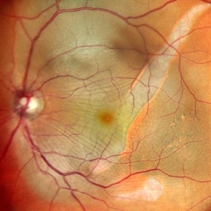 RPE rip in a case of Idiopathic polypoidal choroidopathy
Oct 23 2022 by Anjana Mirajkar, MS Ophthalmology
Color photo central image in a of 61 year old male with RPE rip in a case of Idiopathic Polypoidal Choroidopathy.
Photographer: Dr. Anjana Mirajkar -Retina Foundation, Ahmedabad
Condition/keywords: idiopathic polypoidal choroidopathy, RPE Rip
-
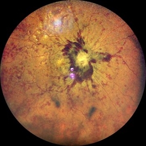
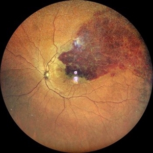
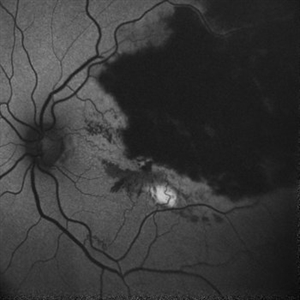
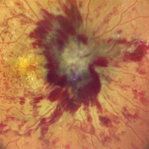
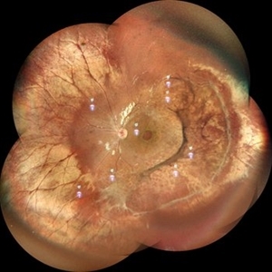
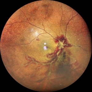
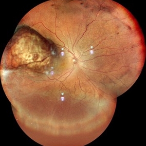
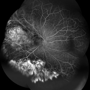
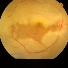
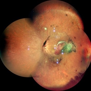
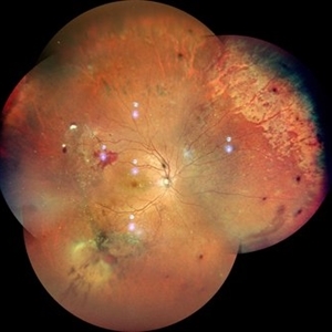
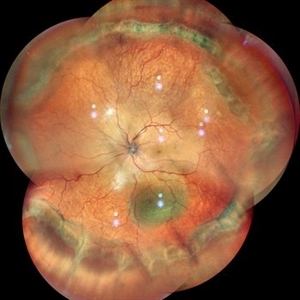
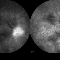
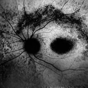
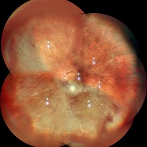
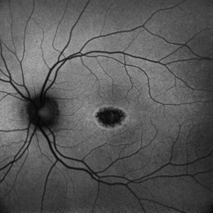
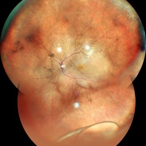
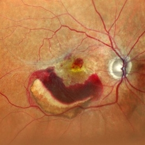
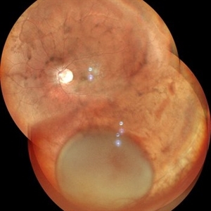
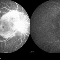
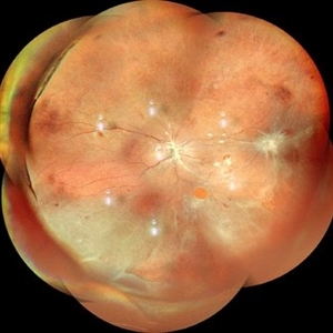
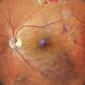
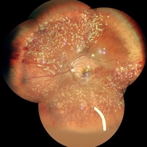
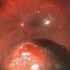
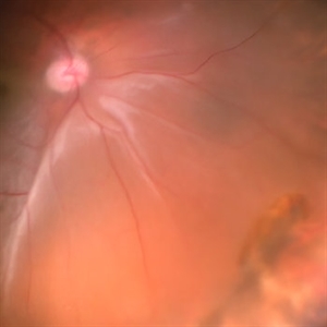
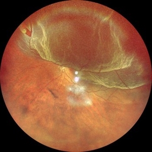
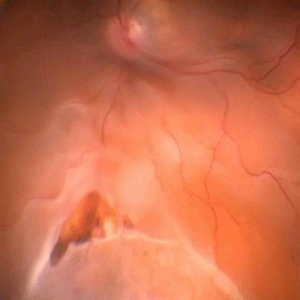
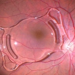
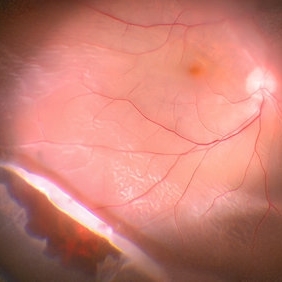
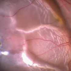
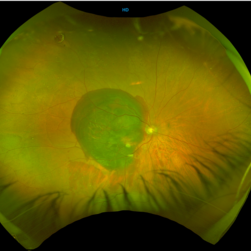
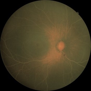
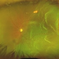
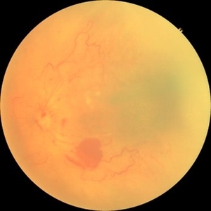
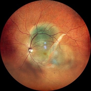 RPE rip in a case of Idiopathic polypoidal choroidopathy
Oct 23 2022 by Anjana Mirajkar, MS Ophthalmology
Color photo wide field image in a of 61 year old male with RPE rip in a case of Idiopathic Polypoidal Choroidopathy.
Photographer: Dr. Anjana Mirajkar -Retina Foundation, Ahmedabad
Condition/keywords: RPE Rip
-
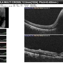
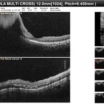
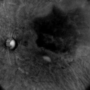
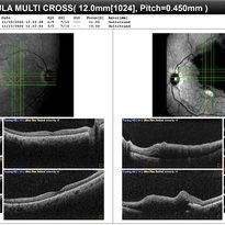
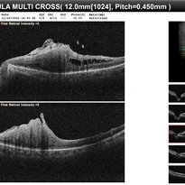
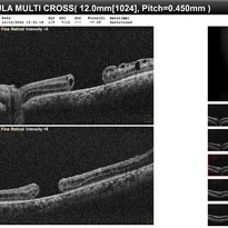
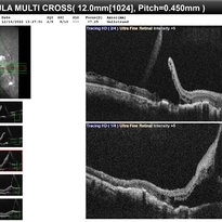
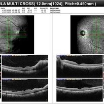
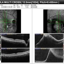
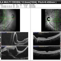
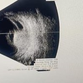
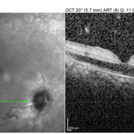
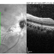
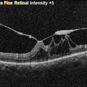
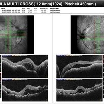
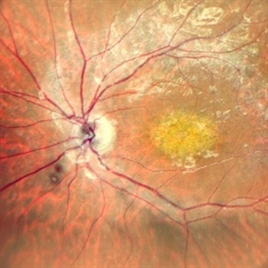
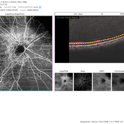
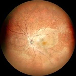
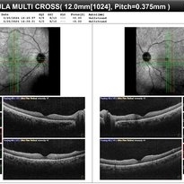
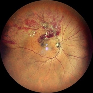
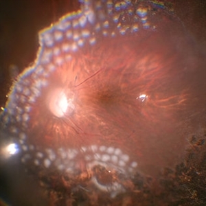
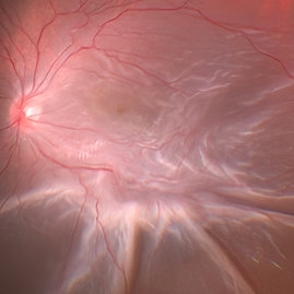
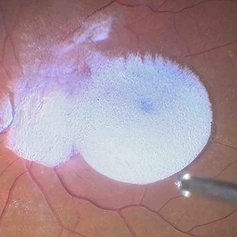
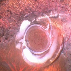
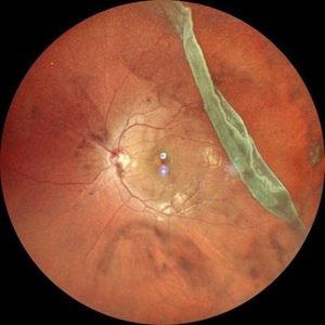
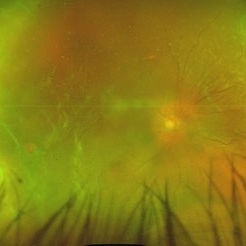
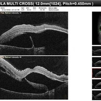 RPE rip in a case of Idiopathic polypoidal choroidopathy
Oct 23 2022 by Anjana Mirajkar, MS Ophthalmology
OCT image in a of 61 year old male with RPE rip in a case of Idiopathic Polypoidal Choroidopathy.
Photographer: Dr. Anjana Mirajkar -Retina Foundation, Ahmedabad
Condition/keywords: Idiopathic polypoidal choroidopathy, RPE rip
-
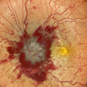
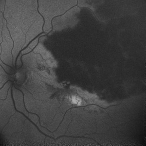
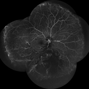
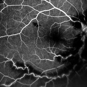
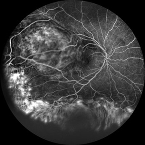
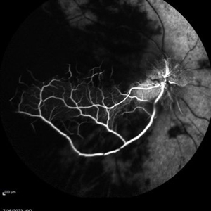
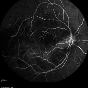
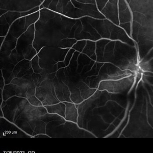
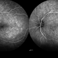
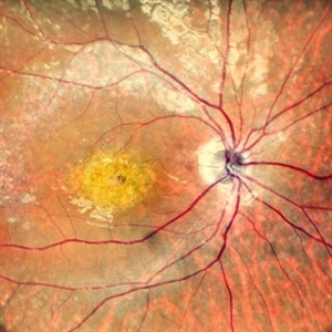
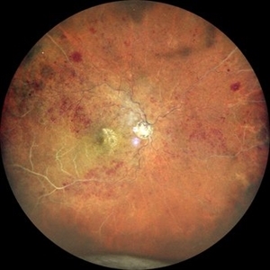
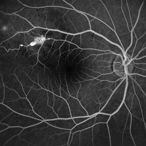
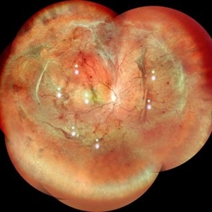
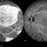
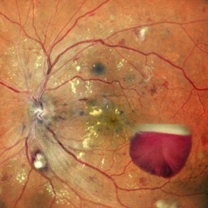
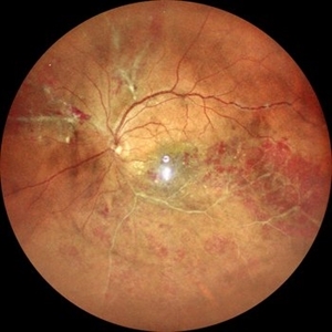
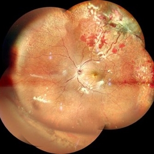
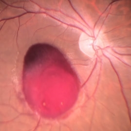
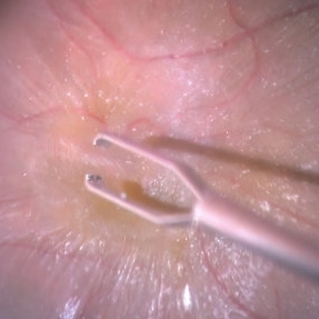
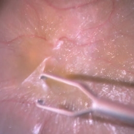
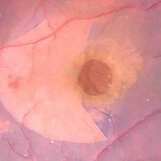
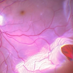
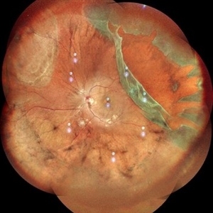
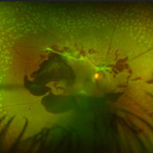
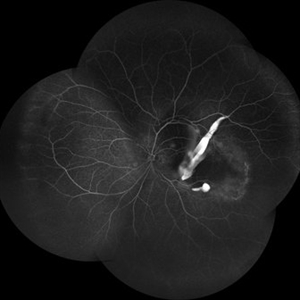 RPE rip in a case of Idiopathic polypoidal choroidopathy
Oct 23 2022 by Anjana Mirajkar, MS Ophthalmology
Montage of Fluorescein angiography in a of 61 year old male with RPE rip in a case of Idiopathic Polypoidal Choroidopathy.
Photographer: Dr. Anjana Mirajkar -Retina Foundation, Ahmedabad
Condition/keywords: Idiopathic polypoidal choroidopathy, RPE rip
-
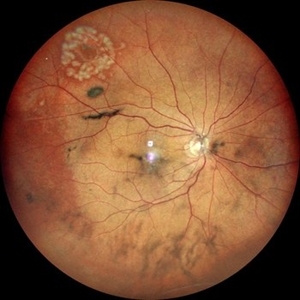
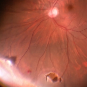
 Idiopathic Intracranial Hypertension
Dec 11 2022 by Anjana Mirajkar, MS Ophthalmology
Wide field color photo of RE of a 23 year old male a case of Idiopathic Intracranial Hypertension
Photographer: Dr. Anjana Mirajkar -Retina Foundation, Ahmedabad
Condition/keywords: benign idiopatic intracranial hypertension
-
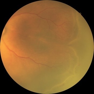
 Idiopathic Intracranial Hypertension
Dec 11 2022 by Anjana Mirajkar, MS Ophthalmology
Central color photo of LE of a 23 year old male a case of Idiopathic Intracranial Hypertension
Photographer: Dr. Anjana Mirajkar -Retina Foundation, Ahmedabad
Condition/keywords: benign idiopatic intracranial hypertension
-
 Idiopathic Intracranial Hypertension
Dec 11 2022 by Anjana Mirajkar, MS Ophthalmology
Wide field color photo of LE of a 23 year old male a case of Idiopathic Intracranial Hypertension
Photographer: Dr. Anjana Mirajkar -Retina Foundation, Ahmedabad
Condition/keywords: benign idiopatic intracranial hypertension
-
 Idiopathic Intracranial Hypertension
Dec 11 2022 by Anjana Mirajkar, MS Ophthalmology
OCT of RE of a 23 year old male a case of Idiopathic Intracranial Hypertension
Photographer: Dr. Anjana Mirajkar -Retina Foundation, Ahmedabad
Condition/keywords: benign idiopatic intracranial hypertension
-
 Idiopathic Intracranial Hypertension
Dec 11 2022 by Anjana Mirajkar, MS Ophthalmology
OCT of LE of a 23 year old male a case of Idiopathic Intracranial Hypertension
Photographer: Dr. Anjana Mirajkar -Retina Foundation, Ahmedabad
Condition/keywords: benign idiopatic intracranial hypertension
-
 Branch Retinal Vein Occlusion with Macular Edema
Dec 11 2022 by Anjana Mirajkar, MS Ophthalmology
Central color photo of a 54 year old male case of LE Branch Retinal Vein Occlusion with Macular Edema.
Photographer: Dr. Anjana Mirajkar -Retina Foundation, Ahmedabad.
Condition/keywords: branch retinal vein occlusion (BRVO)
-
 Branch Retinal Vein Occlusion with Macular Edema.
Dec 11 2022 by Anjana Mirajkar, MS Ophthalmology
Wide field color photo of a 54 year old male case of LE Branch Retinal Vein Occlusion with Macular Edema.
Photographer: Dr. Anjana Mirajkar -Retina Foundation, Ahmedabad.
Condition/keywords: branch retinal vein occlusion (BRVO)
-
 Branch Retinal Vein Occlusion with Macular Edema
Dec 11 2022 by Anjana Mirajkar, MS Ophthalmology
Autofluorescent Image(B-mode) of a 54 year old male case of LE Branch Retinal Vein Occlusion with Macular Edema.
Photographer: Dr. Anjana Mirajkar -Retina Foundation, Ahmedabad
Condition/keywords: branch retinal vein occlusion (BRVO)
-
 Branch Retinal Vein Occlusion with Macular Edema.
Dec 11 2022 by Anjana Mirajkar, MS Ophthalmology
Autofluorescent Image(G-mode) of a 54 year old male case of LE Branch Retinal Vein Occlusion with Macular Edema.
Photographer: Dr. Anjana Mirajkar -Retina Foundation, Ahmedabad
Condition/keywords: branch retinal vein occlusion (BRVO)
-
 Branch Retinal Vein Occlusion with Macular Edema.
Dec 11 2022 by Anjana Mirajkar, MS Ophthalmology
Retro Image of a 54 year old male case of LE Branch Retinal Vein Occlusion with Macular Edema.
Photographer: Dr. Anjana Mirajkar -Retina Foundation, Ahmedabad
Condition/keywords: branch retinal vein occlusion (BRVO)
-
 Branch Retinal Vein Occlusion Macular Edema.
Dec 11 2022 by Anjana Mirajkar, MS Ophthalmology
Retro Image of a 54 year old male case of LE Branch Retinal Vein Occlusion with Macular Edema.
Photographer: Dr. Anjana Mirajkar -Retina Foundation, Ahmedabad
Condition/keywords: branch retinal vein occlusion (BRVO)
-
 Branch Retinal Vein Occlusion with Macular Edema
Dec 11 2022 by Anjana Mirajkar, MS Ophthalmology
OCT of a 54 year old male case of LE Branch Retinal Vein Occlusion with Cystoid Macular Edema.
Photographer: Dr. Anjana Mirajkar -Retina Foundation, Ahmedabad
Condition/keywords: branch retinal vein occlusion (BRVO)
-
 Epiretinal Membrane causing Macular Pucker.
Dec 11 2022 by Anjana Mirajkar, MS Ophthalmology
Color Photo of LE in a 65 year old male case of Epiretinal Membrane causing Macular Pucker.
Photographer: Dr. Anjana Mirajkar -Retina Foundation, Ahmedabad.
Condition/keywords: epiretinal membrane (ERM), macular pucker
-
 Epiretinal Membrane causing Macular Pucker.
Dec 11 2022 by Anjana Mirajkar, MS Ophthalmology
OCT of LE in a 65 year old male case of Epiretinal Membrane causing Macular Pucker.
Photographer: Dr. Anjana Mirajkar -Retina Foundation, Ahmedabad.
Condition/keywords: epiretinal membrane (ERM), macular pucker
-
 Idiopathic Intracranial Hypertension
Dec 11 2022 by Anjana Mirajkar, MS Ophthalmology
Central colour photo of RE of a 23 year old male case of Idiopathic Intracranial Hypertension
Photographer: Dr. Anjana Mirajkar -Retina Foundation, Ahmedabad
Condition/keywords: benign idiopatic intracranial hypertension
-
 FEVR
FEVR
Jan 28 2023 by Anjana Mirajkar, MS Ophthalmology
Colour picture( montage) of RE in a 10 year male child a case of FEVR. with macular hole.
Photographer: Dr. Anjana Mirajkar -Retina Foundation, Ahmedabad.
Condition/keywords: familial exudative vitreoretinopathy (FEVR), macular hole
-
 FEVR
FEVR
Jan 28 2023 by Anjana Mirajkar, MS Ophthalmology
Colour picture( montage) of LE in a 10 year male child a case of FEVR. with macular hole.
Photographer: Dr. Anjana Mirajkar -Retina Foundation, Ahmedabad
Condition/keywords: familial exudative vitreoretinopathy (FEVR), macular hole
-
 Familial Exudative Vitreoretinopathy
Jan 28 2023 by Anjana Mirajkar, MS Ophthalmology
OCT image of LE of 10 year old male child case of FEVR with macular hole.
Photographer: Dr. Anjana Mirajkar -Retina Foundation, Ahmedabad
Condition/keywords: familial exudative vitreoretinopathy (FEVR)
-
 Familial Exudative Vitreoretinopathy
Jan 28 2023 by Anjana Mirajkar, MS Ophthalmology
OCT image of RE of 10 year old male child case of FEVR.
Photographer: Dr. Anjana Mirajkar -Retina Foundation, Ahmedabad
Condition/keywords: familial exudative vitreoretinopathy (FEVR)
-
 Occlusive Vasculitis
Jan 28 2023 by Anjana Mirajkar, MS Ophthalmology
Central color image of RE of a 40 year old female a case of occlusive retinal vasculitis
Photographer: Dr. Anjana Mirajkar -Retina Foundation, Ahmedabad
Condition/keywords: occlusive retinal vasculitis
-
 Occlusive Vasculitis
Jan 28 2023 by Anjana Mirajkar, MS Ophthalmology
Widefield color image of RE of a 40 year old female a case of occlusive retinal vasculitis.
Photographer: Dr. Anjana Mirajkar -Retina Foundation, Ahmedabad
Condition/keywords: occlusive retinal vasculitis
-
 Occlusive Vasculitis
Jan 28 2023 by Anjana Mirajkar, MS Ophthalmology
Wide field FA image of RE of a 40 year old female a case of occlusive retinal vasculitis.
Photographer: Dr. Anjana Mirajkar -Retina Foundation, Ahmedabad
Condition/keywords: occlusive retinal vasculitis
-
 Occlusive Vasculitis
Jan 28 2023 by Anjana Mirajkar, MS Ophthalmology
OCT image of BE of a 40 year old female a case of occlusive retinal vasculitis.
Photographer: Dr. Anjana Mirajkar -Retina Foundation, Ahmedabad
Condition/keywords: occlusive retinal vasculitis
-
 Occlusive Vasculitis
Jan 28 2023 by Anjana Mirajkar, MS Ophthalmology
Central FA picture of a 40 year old female a case of occlusive retinal vasculitis.
Photographer: Dr. Anjana Mirajkar -Retina Foundation, Ahmedabad.
Condition/keywords: occlusive retinal vasculitis
-
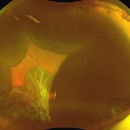
 Choroidal Abscess with occlusive retinal vasculitis
Jan 28 2023 by Anjana Mirajkar, MS Ophthalmology
Widefield color image of LE of a 55 year old male a case of Choroidal abscess with occlusive retinal vasculitis.
Photographer: Dr. Anjana Mirajkar -Retina Foundation, Ahmedabad
Condition/keywords: choroidal abscess
-
 Choroidal Abscess with occlusive retinal vasculitis
Jan 28 2023 by Anjana Mirajkar, MS Ophthalmology
OCT BE of a 55 year old male a case of Choroidal abscess with occlusive retinal vasculitis.
Photographer: Dr. Anjana Mirajkar -Retina Foundation, Ahmedabad
Condition/keywords: Choroidal abscess
-
 Choroidal Hemangioma
Jan 29 2023 by Anjana Mirajkar, MS Ophthalmology
Widefield color image (montage) of RE of a 25 year old female with RE exudative retinal detachment with subretinal mass most likely a Choroidal Hemangioma with fronds of vessels noted inferiorly.
Photographer: Dr. Anjana Mirajkar -Retina Foundation, Ahmedabad
Condition/keywords: choroidal hemangioma
-
 Choroidal Hemangioma
Jan 29 2023 by Anjana Mirajkar, MS Ophthalmology
Widefield color image of RE of a 25 year old female with RE exudative retinal detachment with subretinal mass most likely a Choroidal Hemangioma with fronds of vessels noted inferiorly.
Photographer: Dr. Anjana Mirajkar -Retina Foundation, Ahmedabad
Condition/keywords: choroidal hemangioma
-
 Choroidal Hemangioma
Jan 29 2023 by Anjana Mirajkar, MS Ophthalmology
Widefield fluorescein angiography image (montage) of RE of a 25 year old female with RE exudative retinal detachment with subretinal mass most likely a Choroidal Hemangioma.
Photographer: Dr. Anjana Mirajkar -Retina Foundation, Ahmedabad
Condition/keywords: choroidal hemangioma
-
 Choroidal Hemangioma
Jan 29 2023 by Anjana Mirajkar, MS Ophthalmology
Widefield fluorescein angiography image of RE of a 25 year old female with RE exudative retinal detachment with subretinal mass most likely a Choroidal Hemangioma.
Photographer: Dr. Anjana Mirajkar -Retina Foundation, Ahmedabad
Condition/keywords: choroidal hemangioma
-
 Choroidal Hemangioma
Jan 29 2023 by Anjana Mirajkar, MS Ophthalmology
OCT BE of RE of a 25 year old female with RE exudative retinal detachment with subretinal mass most likely a Choroidal Hemangioma.
Photographer: Dr. Anjana Mirajkar -Retina Foundation, Ahmedabad
Condition/keywords: choroidal hemangioma
-
 Choroidal Hemangioma
Jan 29 2023 by Anjana Mirajkar, MS Ophthalmology
USG BSCAN of RE of a 25 year old female with RE exudative retinal detachment with subretinal mass most likely a Choroidal Hemangioma.
Photographer: Dr. Anjana Mirajkar -Retina Foundation, Ahmedabad
Condition/keywords: unilateral exudative retinal detachment
-
 Central Artery Occlusion
Aug 6 2023 by Anjana Mirajkar, MS Ophthalmology
Color photo of a 42 year old male in a case of central artery occlusion with cilio retinal artery sparing
Photographer: Dr. Anjana Mirajkar -Retina Foundation, Ahmedabad
Condition/keywords: Central Retinal Artery Occlusion, central retinal artery occlusion (CRAO)
-
 Central Artery Occlusion with Cilio Retinal Artery Sparing
Aug 6 2023 by Anjana Mirajkar, MS Ophthalmology
Wide field view of FA (Early phase) of a 42 year old male in a case of central artery occlusion with cilio retinal artery sparing showing delayed arterial filling with choroidal filling
Photographer: Dr. Anjana Mirajkar -Retina Foundation, Ahmedabad
Condition/keywords: central retinal artery occlusion
-
 Central Artery Occlusion with Cilio Retinal Artery Sparing
Aug 6 2023 by Anjana Mirajkar, MS Ophthalmology
Wide field view of FA (Late phase) of a 42 year old male in a case of central artery occlusion with cilio retinal artery sparing showing delayed arterial filling with choroidal filling
Photographer: Dr. Anjana Mirajkar -Retina Foundation, Ahmedabad
Condition/keywords: central retinal artery occlusion (CRAO)
-
 Central Artery Occlusion with Cilio Retinal Artery Sparing
Aug 6 2023 by Anjana Mirajkar, MS Ophthalmology
Central FA frame (Late phase) of a 42 year old male in a case of central artery occlusion with cilio retinal artery sparing
Photographer: Dr. Anjana Mirajkar -Retina Foundation, Ahmedabad
Condition/keywords: central retinal artery occlusion (CRAO)
-
 Central Artery Occlusion with Cilio Retinal Artery Sparing
Aug 6 2023 by Anjana Mirajkar, MS Ophthalmology
OCT image (horizontal scan ) of RE of a 42 year old male in a case of central artery occlusion with cilio retinal artery sparing loss of differentiation of retinal layers in nasal half.
Photographer: Dr. Anjana Mirajkar -Retina Foundation, Ahmedabad
Condition/keywords: Central Retinal Artery Occlusion
-
 Central Artery Occlusion with Cilio Retinal Artery Sparing
Aug 6 2023 by Anjana Mirajkar, MS Ophthalmology
OCT image (Vertical scan ) of RE of a 42 year old male in a case of central artery occlusion with cilio retinal artery sparing showing loss of differentiation of retinal layers in nasal half.
Photographer: Dr. Anjana Mirajkar -Retina Foundation, Ahmedabad
Condition/keywords: central retinal artery occlusion (CRAO)
-
 Superior Retinal Detachment
Aug 13 2023 by Anjana Mirajkar, MS Ophthalmology
A color photo of RE of a 55 year old male in a case of Superior Retinal Detachment with macula off with a superior horse shoe tear with vitreous Haze
Photographer: Dr. Anjana Mirajkar -Retina Foundation, Ahmedabad
Condition/keywords: chronic retinal detachment
-
 Old BRVO with vitreomacular traction
Sep 1 2023 by Anjana Mirajkar, MS Ophthalmology
A fundus image of a 62 year old female case of Old BRVO with traction at the fovea status post laser with anti-VEGF
Photographer: Dr. Anjana Mirajkar -Retina Foundation, Ahmedabad
Condition/keywords: branch retinal vein occlusion (BRVO)
-
 Old BRVO with vitreomacular traction
Sep 1 2023 by Anjana Mirajkar, MS Ophthalmology
A OCT image of a 62 year old female case of Old BRVO with traction at the fovea status post laser with anti-VEGF
Photographer: Dr. Anjana Mirajkar -Retina Foundation, Ahmedabad
-
 BE-OCT
Sep 1 2023 by Anjana Mirajkar, MS Ophthalmology
OCT image of BE of a 30 year old female case of BE Proliferative diabetic retinopathy showing RE-Foveal contour maintained, LE-Foveal contour altered
Photographer: Dr. Anjana Mirajkar -Retina Foundation, Ahmedabad
Condition/keywords: Proliferative Diabetic retinopathy
-
 LE-Sub-hyaloid hemorrhage
Sep 1 2023 by Anjana Mirajkar, MS Ophthalmology
A widefield montage fundus image (LE) of a 30 year old female case of sub hyaloid hemorrhage with vitreous hemorrhage with asteroid hyalosis in case of proliferative diabetic retinopathy
Photographer: Dr. Anjana Mirajkar -Retina Foundation, Ahmedabad
Condition/keywords: proliferative diabetic retinopathy (PDR)
-
 RE- Pre retinal hemorrhage with vitreous hemorrhage
Sep 1 2023 by Anjana Mirajkar, MS Ophthalmology
A widefield montage fundus image (RE) of a 30 year old female case of sub hyaloid hemorrhage with vitreous hemorrhage with asteroid hyalosis in case of proliferative diabetic retinopathy
Photographer: Dr. Anjana Mirajkar -Retina Foundation, Ahmedabad
Condition/keywords: proliferative diabetic retinopathy (PDR)
-
 LE-Sub-hyaloid hemorrhage
Sep 1 2023 by Anjana Mirajkar, MS Ophthalmology
A central fundus image (LE) of a 30 year old female case of sub hyaloid hemorrhage in case of proliferative diabetic retinopathy
Photographer: Dr. Anjana Mirajkar -Retina Foundation, Ahmedabad
Condition/keywords: Proliferative Diabetic retinopathy
-
 LE-Sub-hyaloid hemorrhage
Sep 1 2023 by Anjana Mirajkar, MS Ophthalmology
A wide field fundus image (LE) of a 30 year old female case of sub hyaloid hemorrhage in case of proliferative diabetic retinopathy
Photographer: Dr. Anjana Mirajkar -Retina Foundation, Ahmedabad
Condition/keywords: proliferative diabetic retinopathy (PDR)
-
 VKH
VKH
Sep 29 2023 by Anjana Mirajkar, MS Ophthalmology
Wide field color photo image of LE of a 41 year old female case of VKH showing multiple fluid pockets in the posterior pole with ILM folds
Photographer: Dr. Anjana Mirajkar -Retina Foundation, Ahmedabad
Imaging device: Mirante-Nidek
Condition/keywords: vkh
-
 VKH
VKH
Sep 29 2023 by Anjana Mirajkar, MS Ophthalmology
Wide field color photo image of RE of a 41 year old female case of VKH showing exudative retinal detachment inferiorly with multiple fluid pockets in the posterior pole with ILM folds
Photographer: Dr. Anjana Mirajkar -Retina Foundation, Ahmedabad
Imaging device: Mirante-Nidek
Condition/keywords: Vogt-Koyanagi-Harada
-
 VKH
VKH
Sep 29 2023 by Anjana Mirajkar, MS Ophthalmology
OCT of BE of a 41 year old female in a case of BE VKH showing subretinal fluid with septations with choroidal undulations.
Photographer: Dr. Anjana Mirajkar -Retina Foundation, Ahmedabad
Imaging device: Mirante-Nidek
Condition/keywords: vkh, Vogt-Koyanagi-Harada
-
 VKH
VKH
Sep 29 2023 by Anjana Mirajkar, MS Ophthalmology
Late frame of FA+ICG of LE of a 41 year old female showing disc leakage with hyperfluorescence suggestive of leakage with hypofluoroscence on FA and ICG in a case of VKH.
Photographer: Dr. Anjana Mirajkar -Retina Foundation, Ahmedabad.
Imaging device: Heidelberg
Condition/keywords: vkh
-
 VKH
VKH
Sep 29 2023 by Anjana Mirajkar, MS Ophthalmology
Late frame of FA+ICG of RE of a 41 year old female showing disc leakage with hyperfluorescence suggestive of leakage with hypofluoroscence on FA and ICG in a case of VKH.
Photographer: Dr. Anjana Mirajkar -Retina Foundation, Ahmedabad.
Imaging device: Heidelberg
Condition/keywords: Vogt-Koyanagi-Harada
-
 Macular Dystrophy
Nov 3 2023 by Anjana Mirajkar, MS Ophthalmology
An autofluorescence image -OD of a 22 year old male case of macular dystrophy showing hypo autofluorescence
Photographer: Dr. Anjana Mirajkar -Retina Foundation, Ahmedabad
Imaging device: Mirante-Nidek
Condition/keywords: heredomacular degeneration, Macular Dystrophy
-
 Macular Dystrophy
Nov 3 2023 by Anjana Mirajkar, MS Ophthalmology
An autofluorescence image -OS of a 22 year old male case of macular dystrophy showing hypo autofluorescence
Photographer: Dr. Anjana Mirajkar -Retina Foundation, Ahmedabad
Imaging device: Mirante-Nidek
Condition/keywords: heredomacular degeneration, Macular Dystrophy
-
 Macular Dystrophy
Nov 3 2023 by Anjana Mirajkar, MS Ophthalmology
A central color photo OS of a 22 year old male case of macular dystrophy.
Photographer: Dr. Anjana Mirajkar -Retina Foundation, Ahmedabad
Imaging device: Mirante-Nidek
Condition/keywords: heredomacular degeneration, Macular Dystrophy
-
 Macular Dystrophy
Nov 3 2023 by Anjana Mirajkar, MS Ophthalmology
A central color photo OD of a 22 year old male case of macular dystrophy.
Photographer: Dr. Anjana Mirajkar -Retina Foundation, Ahmedabad
Imaging device: Mirante-Nidek
Condition/keywords: heredomacular degeneration, Macular Dystrophy
-
 Retinal Detachment
Nov 3 2023 by Anjana Mirajkar, MS Ophthalmology
A widefield image of OS of a 55 year old female case of inferior retinal detachment with macula off.
Photographer: Dr. Anjana Mirajkar -Retina Foundation, Ahmedabad
Imaging device: Mirante-Nidek
Condition/keywords: inferior retinal detachment, Retinal Detachment
-
 Retinal Detachment
Nov 3 2023 by Anjana Mirajkar, MS Ophthalmology
A widefield image (montage) of OS of a 55 year old female case of inferior retinal detachment with macula off.
Photographer: Dr. Anjana Mirajkar -Retina Foundation, Ahmedabad
Imaging device: Mirante-Nidek
Condition/keywords: inferior retinal detachment, Retinal Detachment
-
 Central vein occlusion (chronic)
Nov 26 2023 by Anjana Mirajkar, MS Ophthalmology
A widefield color photo image of RE of a 60 year old male in a case of central vein occlusion.
Photographer: Dr. Anjana Mirajkar -Retina Foundation, Ahmedabad
Imaging device: Mirante-Nidek
Condition/keywords: central retinal vein occlusion (CRVO), central vein occlusion, ischemic CRVO
-
 Central vein occlusion (chronic)
Nov 26 2023 by Anjana Mirajkar, MS Ophthalmology
A central color photo image of RE of a 60 year old male in a case of central vein occlusion.
Photographer: Dr. Anjana Mirajkar -Retina Foundation, Ahmedabad
Imaging device: Mirante-Nidek
Condition/keywords: central retinal vein occlusion (CRVO), ischemic CRVO
-
 Heredomacular degeneration
Nov 30 2023 by Anjana Mirajkar, MS Ophthalmology
Central autofluorescence image of RE of an 30 year old male in case of heredo-macular degeneration showing hypo fluorescence.
Photographer: Dr. Anjana Mirajkar -Retina Foundation, Ahmedabad
Imaging device: Mirante-Nidek
Condition/keywords: heredomacular degeneration, HMD
-
 Heredomacular degeneration
Nov 30 2023 by Anjana Mirajkar, MS Ophthalmology
Central autofluorescence image of LE of an 30 year old male in case of heredo-macular degeneration showing hypo fluorescence.
Photographer: Dr. Anjana Mirajkar -Retina Foundation, Ahmedabad
Imaging device: Mirante-Nidek
Condition/keywords: heredomacular degeneration, HMD
-
 Asteroid hyalosis
Dec 1 2023 by Anjana Mirajkar, MS Ophthalmology
A widefield image of RE of a 65 year old male with dense asteroid Hyalosis
Photographer: Dr. Anjana Mirajkar -Retina Foundation, Ahmedabad
Imaging device: Mirante-Nidek
Condition/keywords: asteroid hyalosis, vitreous degeneration
-
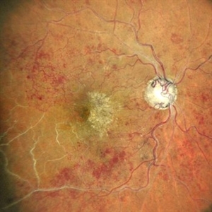
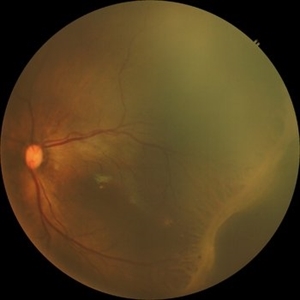
 Proliferative diabetic retinopathy
Jan 28 2024 by Anjana Mirajkar, MS Ophthalmology
A central fundus image of a 60 year old male showing neovascularization at the disc in a case of PDR.
Photographer: Dr. Anjana Mirajkar -Retina Foundation, Ahmedabad
Imaging device: RS1-Nidek
Condition/keywords: Neovascularisation at the Disc (NVD), proliferative diabetic retinopathy (PDR)
-
 Proliferative diabetic retinopathy
Jan 28 2024 by Anjana Mirajkar, MS Ophthalmology
An OCT-A image of a 60 year old male showing neovascularisation at the disc in a case of PDR.
Photographer: Dr. Anjana Mirajkar -Retina Foundation, Ahmedabad
Imaging device: RS1-nidek
Condition/keywords: Neovascularisation at the Disc (NVD), proliferative diabetic retinopathy (PDR)
-
 Dislocated IOL
Jan 28 2024 by Anjana Mirajkar, MS Ophthalmology
A widefield image of a 60 year old female with dislocated IOL inferiorly.
Photographer: Dr. Anjana Mirajkar -Retina Foundation, Ahmedabad
Imaging device: Mirante-Nidek
Condition/keywords: dislocated intraocular lens (IOL)
-
 Macroaneurysms
Jan 28 2024 by Anjana Mirajkar, MS Ophthalmology
Fundus image in a 20 year old female showing multiple macro aneurysms surrounded with exudation along the supero-temporal arcade and hard exudates at macula suggestive of macular edema.
Photographer: Dr. Anjana Mirajkar -Retina Foundation, Ahmedabad
Imaging device: Mirante-Nidek
Condition/keywords: macroaneurysm
-
 Macroaneurysms
Jan 28 2024 by Anjana Mirajkar, MS Ophthalmology
Fundus image of post focal laser in a 20 year old female in a case of retinal arterial macroaneurysm
Photographer: Dr. Anjana Mirajkar -Retina Foundation, Ahmedabad
Imaging device: Mirante-Nidek
Condition/keywords: macroarterial aneurysm
-
 Macroaneurysms
Jan 28 2024 by Anjana Mirajkar, MS Ophthalmology
Fundus fluorescein angiography image (Late phase) in a 20 year old female showing leakage in a case of retinal artery macro-aneurysms.
Photographer: Dr. Anjana Mirajkar -Retina Foundation, Ahmedabad
Imaging device: Mirante-Nidek
Condition/keywords: retinal arterial macroaneurysm
-
 macular hole
Jan 28 2024 by Anjana Mirajkar, MS Ophthalmology
Fundus photograph of a 40 year old female showing macular hole with 2 retinal holes surrounding it
Photographer: Dr. Anjana Mirajkar -Retina Foundation, Ahmedabad
Imaging device: Mirante-Nidek
Condition/keywords: macular hole, multiple retinal holes
-
 Wet age related macular degeneration
Jan 28 2024 by Anjana Mirajkar, MS Ophthalmology
Fundus photograph of an 70 year old male with sub retinal bleed and exudation as well as scarring in case of wet age related macular degeneration.
Photographer: Dr. Anjana Mirajkar -Retina Foundation, Ahmedabad
Imaging device: Mirante-Nidek
Condition/keywords: choroidal neovascular membrane (CNVM), wet age-related macular degeneration (wet AMD)
-
 Retinal Hole
Feb 11 2024 by Anjana Mirajkar, MS Ophthalmology
A color photo of RE of a 50 year old male showing lasered retinal hole superiorly with vitreous degeneration.
Photographer: Dr. Anjana Mirajkar -Retina Foundation, Ahmedabad
Imaging device: Mirante-Nidek
Condition/keywords: full thickness retinal hole
-
 Retinal Hole
Feb 11 2024 by Anjana Mirajkar, MS Ophthalmology
A color photo of RE of a 50 year old male showing retinal hole superiorly with vitreous degeneration.
Photographer: Dr. Anjana Mirajkar -Retina Foundation, Ahmedabad
Imaging device: Mirante-Nidek
Condition/keywords: retinal hole
-
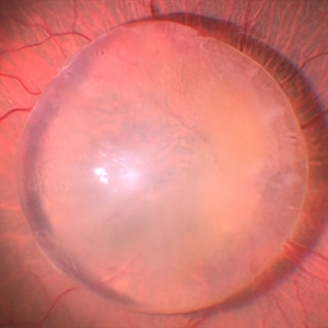
 Dislocated Cataractous Lens
Feb 11 2024 by Anjana Mirajkar, MS Ophthalmology
A wide field image of LE of a 40 year old male showing inferior dislocation of crystalline lens which is cataractous in vitreous cavity.
Photographer: Dr. Anjana Mirajkar -Retina Foundation, Ahmedabad
Imaging device: Mirante-Nidek
Condition/keywords: dislocated crystalline lens
-
 Tractional Retinal Detachment
Mar 22 2024 by Anjana Mirajkar, MS Ophthalmology
A widefield color photo of RE of a 17 year old male showing tractional retinal detachment likely a ROP sequelae.
Photographer: Dr. Anjana Mirajkar -Retina Foundation, Ahmedabad
Imaging device: Mirante-Nidek
Condition/keywords: ROP sequelae
-
 Syphilitic Posterior Uveitis
Mar 22 2024 by Anjana Mirajkar, MS Ophthalmology
A color photo image of LE of a 36 year old female showing hypopigmented lesions at the posterior pole(ground glass appearance) in a case of syphilitic posterior placoid chorioretinitis
Photographer: Dr. Anjana Mirajkar -Retina Foundation, Ahmedabad
Imaging device: Mirante-Nidek
Condition/keywords: syphilitic posterior uveitis
-
 Syphilitic Posterior Uveitis
Mar 22 2024 by Anjana Mirajkar, MS Ophthalmology
A color photo image of RE of a 36 year old female showing hypopigmented lesions at the posterior pole(ground glass appearance) in a case of syphilitic posterior placoid chorioretinitis
Photographer: Dr. Anjana Mirajkar -Retina Foundation, Ahmedabad
Imaging device: Mirante-Nidek
Condition/keywords: posterior uveitis
-
 Syphilitic Posterior Uveitis
Mar 22 2024 by Anjana Mirajkar, MS Ophthalmology
FA image of LE of a 36 year old female showing hyper-fluorescence (staining) from early to late phases of the angiogram in a case syphilitic posterior placoid chorioretinitis. ICG image depicts hypo-cyanence from early to late phases.
Photographer: Dr. Anjana Mirajkar -Retina Foundation, Ahmedabad
Condition/keywords: acute syphilitic posterior placoid chorioretinitis
-
 Syphilitic Posterior Uveitis
Mar 22 2024 by Anjana Mirajkar, MS Ophthalmology
FA image of RE of a 36 year old female showing hyper-fluorescence (staining) from early to late phases of the angiogram in a case syphilitic posterior placoid chorioretinitis. ICG image depicts hypo-cyanence from early to late phases.
Photographer: Dr. Anjana Mirajkar -Retina Foundation, Ahmedabad
Imaging device: Heidelberg
Condition/keywords: acute syphilitic posterior placoid chorioretinitis
-
 Syphilitic Posterior Uveitis
Mar 22 2024 by Anjana Mirajkar, MS Ophthalmology
An OCT image BE of 36 year old female showing RPE granularity and IS/OS irregularity in a case of syphilitic posterior placoid chorioretinitis
Photographer: Dr. Anjana Mirajkar -Retina Foundation, Ahmedabad
Condition/keywords: acute posterior placoid chorioretinitis, acute syphilitic posterior placoid chorioretinitis
-
 Proliferative diabetic retinopathy
Apr 28 2024 by Anjana Mirajkar, MS Ophthalmology
A widefield color image of a 60 year old male with type II diabetes showing sub hyaloid hemorrhage with traction at the fovea with hard exudates with venous looping along the supero temporal arcade with NVE inferiorly with surrounding laser marks.
Photographer: Dr. Anjana Mirajkar -Retina Foundation, Ahmedabad
Imaging device: Mirante-Nidek
Condition/keywords: pan-retinal photocoagulation (PRP), proliferative diabetic retinopathy (PDR)
-
 Retinal Detachment
Apr 28 2024 by Anjana Mirajkar, MS Ophthalmology
A montage of a 40 year old male showing multiple breaks with inferior retinal detachment with peripheral traction in a silicon filled eye.
Photographer: Dr. Anjana Mirajkar -Retina Foundation, Ahmedabad
Imaging device: Mirante-Nidek
Condition/keywords: Retinal detachment under Silicon Oil
-
 Branch Retinal Vein Occlusion
Apr 28 2024 by Anjana Mirajkar, MS Ophthalmology
A widefield color photo of a 55 year old male case of supero-temporal BRVO showing venous tortuosity, cotton wool spots, flame shaped hemorrhages and macular edema.
Photographer: Dr. Anjana Mirajkar -Retina Foundation, Ahmedabad
Imaging device: Mirante-Nidek
Condition/keywords: branch retinal vein occlusion (BRVO), ST BRVO
-
 Proliferative Diabetic Retinopathy
May 24 2024 by Anjana Mirajkar, MS Ophthalmology
A central photo of a 50 year old male case of PDR showing a sub-hyaloid hemorrhage with cotton wool spots , hard exudates at the fovea with dot and blot hemorrhages.
Photographer: Dr. Anjana Mirajkar -Retina Foundation, Ahmedabad
Imaging device: Mirante-Nidek
Condition/keywords: proliferative diabetic retinopathy (PDR)
-
 Proliferative Diabetic Retinopathy
May 24 2024 by Anjana Mirajkar, MS Ophthalmology
A central photo of a 65 year old female of right eye showing fibro vascular proliferation with neovascularization elsewhere in a case of proliferative diabetic retinopathy.
Photographer: Dr. Anjana Mirajkar -Retina Foundation, Ahmedabad
Imaging device: Mirante-Nidek
Condition/keywords: NVE, proliferative diabetic retinopathy (PDR), tractional retinal detachment
-
 Proliferative Diabetic Retinopathy
May 24 2024 by Anjana Mirajkar, MS Ophthalmology
A central photo of a 60 year old male of left eye case of neovascularization elsewhere with dot and blot hemorrhages in a case of proliferative diabetic retinopathy.
Photographer: Dr. Anjana Mirajkar -Retina Foundation, Ahmedabad
Imaging device: Mirante-Nidek
Condition/keywords: NVE, pre-proliferative diabetic retinopathy
-
 Dislocated IOL
May 24 2024 by Anjana Mirajkar, MS Ophthalmology
Intra-operative photo of a dislocated IOL in the left eye.
Photographer: Dr. Anjana Mirajkar -Retina Foundation, Ahmedabad
Condition/keywords: dislocated IOL, dislocated posterior chamber intraocular lens (PCIOL)
-
 Idiopathic Retinal Vasculitis
Jun 9 2024 by Anjana Mirajkar, MS Ophthalmology
A widefield image of a 32 year old male of LE showing sclerosed vessels more prominent inferiorly with superficial hemorrhages noted in all quadrants along with sheathing of vessels noted in superiorly.
Photographer: Dr. Anjana Mirajkar -Retina Foundation, Ahmedabad
Imaging device: Mirante-Nidek
Condition/keywords: Eales disease
-
 Idiopathic Retinal Vasculitis
Jun 9 2024 by Anjana Mirajkar, MS Ophthalmology
A widefield image of a 32 year old male of LE showing laser marks in inferior and superior half with an floating ozurdex implant (inferiorly) in a case of idiopathic retinal vasculitis.
Photographer: Dr. Anjana Mirajkar -Retina Foundation, Ahmedabad
Imaging device: Mirante-Nidek
Condition/keywords: idiopathic retinal vasculitis, laser photocoagulation, Ozurdex implant, pan-retinal photocoagulation (PRP)
-
 Idiopathic Retinal Vasculitis
Jun 9 2024 by Anjana Mirajkar, MS Ophthalmology
A color photo montage of an 32 year old male of LE showing laser marks in inferior and superior half with an floating ozurdex implant (inferiorly) in a case of idiopathic retinal vasculitis.
Photographer: Dr. Anjana Mirajkar -Retina Foundation, Ahmedabad
Imaging device: Mirante-Nidek
Condition/keywords: idiopathic retinal vasculitis, laser photocoagulation, Ozurdex implant
-
 Von Hippel Lindau Syndrome
Jun 9 2024 by Anjana Mirajkar, MS Ophthalmology
A widefield montage of a 23 year old female of LE case of VHL syndrome showing some hemorrhages with traction superiorly in a silicon oil filled eye with central settled retina. Cryo and laser marks are noted in periphery.
Photographer: Dr. Anjana Mirajkar -Retina Foundation, Ahmedabad
Imaging device: Mirante-Nidek
Condition/keywords: cryotherapy, exudative detachment, laser photocoagulation, vitreous hemorrhage, Von Hippel-Lindau
-
 Laser Done Around Retinectomy
Jun 16 2024 by Anjana Mirajkar, MS Ophthalmology
An intra-operative image of RE showing retinectomy done inferiorly in view of contracted retina which is lasered. Old laser marks can be seen superiorly.
Photographer: Dr. Anjana Mirajkar -Retina Foundation, Ahmedabad
Condition/keywords: Fibrosed retina, retinectomy
-
 Horseshoe Tear
Jun 16 2024 by Anjana Mirajkar, MS Ophthalmology
An intra operative image showing a large horse shoe tear with retinal detachment.
Photographer: Dr. Anjana Mirajkar -Retina Foundation, Ahmedabad
Condition/keywords: rhegmatogenous retinal detachment
-
 Lasered Horseshoe Tear
Jun 16 2024 by Anjana Mirajkar, MS Ophthalmology
An intra operative image showing a lasered horse shoe tear which is being indented for checking the tear being lasered 360 degree.
Photographer: Dr. Anjana Mirajkar -Retina Foundation, Ahmedabad
-
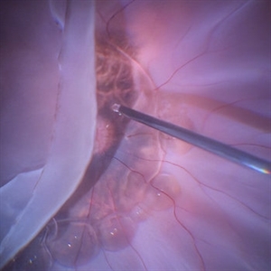
 Retinal Detachment With PVR Changes
Jun 16 2024 by Anjana Mirajkar, MS Ophthalmology
An intra operative still of RE of a 32 year old female case of retinal detachment with Star fold along the supero temporal quadrant.
Photographer: Dr. Anjana Mirajkar -Retina Foundation, Ahmedabad
Condition/keywords: proliferative vitreoretinopathy (PVR)
-
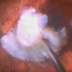
 Dislocated Cataractous Lens
Jun 16 2024 by Anjana Mirajkar, MS Ophthalmology
An intra operative still image of a 65 year old male showing an dislocated cataractous lens during cataract surgery which was removed during vitrectomy and and secondary IOL was placed.
Photographer: Dr. Anjana Mirajkar -Retina Foundation, Ahmedabad
Condition/keywords: dislocated crystalline lens
-
 Dislocated Lens Lying on the Retina
Jul 3 2024 by Anjana Mirajkar, MS Ophthalmology
An intraoperative still showing us an dislocated crystalline lens lying on the retina.
Photographer: Dr. Anjana Mirajkar -Retina Foundation, Ahmedabad
Condition/keywords: dislocated crystalline lens
-
 Dislocated Lens
Jul 3 2024 by Anjana Mirajkar, MS Ophthalmology
An intra operative image showing us the dislocated cataractous lens piece eaten up by the cutter.
Photographer: Dr. Anjana Mirajkar -Retina Foundation, Ahmedabad.
Condition/keywords: Dislocated lens piece eaten up by the cutter
-
 Star Folds in a Chronic Retinal Detachment
Jul 3 2024 by Anjana Mirajkar, MS Ophthalmology
Intra-operative still RE showing a star fold at the parafoveal area causing traction at the macula. Brilliant blue dye being injected to the stain the ILM.
Photographer: Dr. Anjana Mirajkar -Retina Foundation, Ahmedabad
Condition/keywords: brilliant blue staining, proliferative vitreoretinopathy (PVR), star folds
-
 Retinal Detachment With Vitreous Hemorrhage With Sub Retinal Bleed With PVR Changes
Jul 4 2024 by Anjana Mirajkar, MS Ophthalmology
Intra operative image showing us sub retinal bleed at the macular area with superior vitreous hemorrhage with retinal detachment with inferior PVR changes likely in a case of trauma.
Photographer: Dr. Anjana Mirajkar -Retina Foundation, Ahmedabad
Condition/keywords: ocular trauma, retinal detachment with PVR, SUB RETINAL HEMORRHAGE, vitreous hemorrhage
-
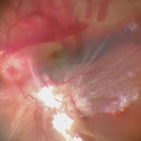
 Retinal Detachment With Retinal Break
Jul 4 2024 by Anjana Mirajkar, MS Ophthalmology
Intra operative image showing us a bullous retinal detachment with retinal break noted inferiorly.
Photographer: Dr. Anjana Mirajkar -Retina Foundation, Ahmedabad
Condition/keywords: retinal hole, rhegmatogenous retinal detachment
-
 ILM Staining in Case of Macular Hole
Jul 4 2024 by Anjana Mirajkar, MS Ophthalmology
Intra operative still of LE showing ILM staining done with BBG dye in case of macular hole.
Photographer: Dr. Anjana Mirajkar -Retina Foundation, Ahmedabad
Condition/keywords: ILM staining, macular hole
-
 Pre retinal Hemorrhage
Jul 5 2024 by Anjana Mirajkar, MS Ophthalmology
An intra operative image of a pre retinal hemorrhage like a sub hyaloid hemorrhage at the macular area.
Photographer: Dr. Anjana Mirajkar -Retina Foundation, Ahmedabad
-
 Retinal Break Inferiorly
Jul 5 2024 by Anjana Mirajkar, MS Ophthalmology
An Intra operative still showing a break inferiorly.
Photographer: Dr. Anjana Mirajkar -Retina Foundation, Ahmedabad
Condition/keywords: retinal break
-
 Removal of Hyaloid
Jul 5 2024 by Anjana Mirajkar, MS Ophthalmology
An intra operative still of hyaloid removal done with forceps.
Photographer: Dr. Anjana Mirajkar -Retina Foundation, Ahmedabad
Condition/keywords: posterior hyaloid
-
 Superior Retinal Detachment With Horse Shoe Tear
Jul 18 2024 by Anjana Mirajkar, MS Ophthalmology
A widefield photo of LE showing superior retinal detachment involving the macula with a horse shoe tear at 11 'o clock
Photographer: Dr. Anjana Mirajkar -Retina Foundation, Ahmedabad
Imaging device: Mirante-Nidek
-
 Membranes Formed Under Silicon Oil and Retina
Jul 18 2024 by Anjana Mirajkar, MS Ophthalmology
A montage color photo of RE of a 7 year old male with membranes formed between silicon and the retina injected for retinal detachment.
Photographer: Dr. Anjana Mirajkar -Retina Foundation, Ahmedabad
Imaging device: Mirante-Nidek
Condition/keywords: sub-silicon membranes
-
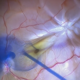
 Epiretinal Membrane
Jul 19 2024 by Anjana Mirajkar, MS Ophthalmology
An intra operative still showing removal of the epi -retinal membrane with forceps under high magnification after staining with triamcinolone acetonide dye.
Photographer: Dr. Anjana Mirajkar -Retina Foundation, Ahmedabad
Imaging device: Mirante-Nidek
Condition/keywords: epiretinal membrane removal
-
 Retinal Artery Macro-aneurysms
Jul 19 2024 by Anjana Mirajkar, MS Ophthalmology
An intra operative still of LE showing a retinal artery macro aneurysm causing a sub hylaoid and sub ILM hemorrhage.
Photographer: Dr. Anjana Mirajkar -Retina Foundation, Ahmedabad
Imaging device: Mirante-Nidek
Condition/keywords: retinal arterial macroaneurysm, sub hyaloid hemorrhage, sub internal limiting membrane haemorrhage
-
 Large Horse Shoe Tear
Jul 19 2024 by Anjana Mirajkar, MS Ophthalmology
An intra operative still showing us a large horse shoe tear which has posterior location inferiorly causing an retina detachment.
Photographer: Dr. Anjana Mirajkar -Retina Foundation, Ahmedabad
Imaging device: Mirante-Nidek
-
 Intravitreal Triamcinolone Acetonide
Jul 19 2024 by Anjana Mirajkar, MS Ophthalmology
An intra operative still showing us triamcinolone acetonide crystals resting on the retina(posterior pole) forming the two halos just before the hyaloid removal.
Photographer: Dr. Anjana Mirajkar -Retina Foundation, Ahmedabad
Imaging device: Mirante-Nidek
Condition/keywords: intravitreal, triamcinolone
-
 ILM Peeling in a Case of Macular Hole
Sep 28 2024 by Anjana Mirajkar, MS Ophthalmology
An intra operative image showing staining of the ILM followed by its peeling done in case of macular hole.
Photographer: Dr. Anjana Mirajkar -Retina Foundation, Ahmedabad
Condition/keywords: ILM peeling, Macular hole
-
 Giant Retinal Tear
Sep 28 2024 by Anjana Mirajkar, MS Ophthalmology
An intra operative image of the right eye showing a giant retinal tear with a superior retinal detachment.
Photographer: Dr. Anjana Mirajkar -Retina Foundation, Ahmedabad
Condition/keywords: GIANT RETINAL TEAR
-
 Dislocated Lens (IOL) Lying on the Retina
Sep 28 2024 by Anjana Mirajkar, MS Ophthalmology
An intraoperative image of right eye showing dislocated IOL with the capsular bag lying on the posterior pole with surrounding pigmentary changes.
Photographer: Dr. Anjana Mirajkar -Retina Foundation, Ahmedabad
Condition/keywords: dislocated IOL
-
 Proliferative Vitreoretinopathy
Sep 28 2024 by Anjana Mirajkar, MS Ophthalmology
An intra operative image of right eye showing multiple star folds involving the macular area.
Photographer: Dr. Anjana Mirajkar -Retina Foundation, Ahmedabad
Condition/keywords: Starfolds
-
 Dislocated IOL
Sep 28 2024 by Anjana Mirajkar, MS Ophthalmology
An intra operative image of right eye showing dislocated IOL sitting on the posterior pole.
Photographer: Dr. Anjana Mirajkar -Retina Foundation, Ahmedabad
Condition/keywords: dislocated IOL
-
 Retinal Detachment With a Horse Shoe Tear
Sep 28 2024 by Anjana Mirajkar, MS Ophthalmology
An intra operative image showing retinal detachment in the left eye with an horse shoe tear at 5 o'clock.
Photographer: Dr. Anjana Mirajkar -Retina Foundation, Ahmedabad
Condition/keywords: rhegmatogenous retinal detachment
-
 Large Subretinal Bleed in Case of Wet ARMD
Sep 28 2024 by Anjana Mirajkar, MS Ophthalmology
An intra operative image showing large sub retinal hemorrhage involving the macular area and along the superior arcade with exudation at the macular area in case of wet ARMD.
Photographer: Dr. Anjana Mirajkar -Retina Foundation, Ahmedabad
Condition/keywords: polypoidal choroidal vasculopathy (PCV), subretinal hemorrhage, wet age-related macular degeneration (wet AMD)
-
 ILM Peeling in Case of Macular Hole
Sep 28 2024 by Anjana Mirajkar, MS Ophthalmology
An intra operative still showing a stained ILM removal done with forceps in case of large macular hole.
Photographer: Dr. Anjana Mirajkar -Retina Foundation, Ahmedabad
Condition/keywords: internal limiting membrane (ILM) peeling, Macular hole
-
 Giant Retinal Tear
Sep 28 2024 by Anjana Mirajkar, MS Ophthalmology
An intra-operative still showing giant retinal tear from 6 to 11 clock hour with folded posterior margins.
Photographer: Dr. Anjana Mirajkar -Retina Foundation, Ahmedabad
Condition/keywords: GIANT RETINAL TEAR
-
 Retinal Detachment With a Retinal Tear Inferiorly
Sep 28 2024 by Anjana Mirajkar, MS Ophthalmology
An intra operative still of the right eye showing retinal detachment with large retinal tear inferiorly.
Photographer: Dr. Anjana Mirajkar -Retina Foundation, Ahmedabad
Condition/keywords: retinal tear
-
 Giant Retinal Tear
Sep 28 2024 by Anjana Mirajkar, MS Ophthalmology
An intra operative image showing slow injection of PFCL on the posterior pole to unfold the posterior margins in case of giant retinal tear.
Photographer: Dr. Anjana Mirajkar -Retina Foundation, Ahmedabad
Condition/keywords: GIANT RETINAL TEAR
-
 Flattening Out the Retina With PFCL
Sep 28 2024 by Anjana Mirajkar, MS Ophthalmology
An intra operative image showing slow injection of PFCL to flatten out the folded retina.
Photographer: Dr. Anjana Mirajkar -Retina Foundation, Ahmedabad
Condition/keywords: GIANT RETINAL TEAR, PFCL
-
 ILM Peeling in a Case of Large Macular Hole
Sep 28 2024 by Anjana Mirajkar, MS Ophthalmology
An intra operative still showing stained ILM peeling done with forceps in a case of large macular hole.
Photographer: Dr. Anjana Mirajkar -Retina Foundation, Ahmedabad
Condition/keywords: full thickness macular hole, internal limiting membrane (ILM) peeling
-
 Retinal Detachment Secondary to Retinal Hole
Sep 28 2024 by Anjana Mirajkar, MS Ophthalmology
An intra operative image showing retinal detachment involving the macula secondary to retinal hole.
Photographer: Dr. Anjana Mirajkar -Retina Foundation, Ahmedabad
Condition/keywords: retinal hole, rhegmatogenous retinal detachment
-
 Giant Retinal Tear
Oct 11 2024 by Anjana Mirajkar, MS Ophthalmology
Widefield fundus photograph of LE showing giant retinal tear extending from 12 to 4 o clock.
Photographer: Dr. Anjana Mirajkar -Retina Foundation, Ahmedabad
Imaging device: Mirante-Nidek
Condition/keywords: giant retinal tear
-
 Giant Retinal Tear
Oct 11 2024 by Anjana Mirajkar, MS Ophthalmology
Fundus photograph montage of LE showing a giant retinal extending from 12 to 4 o clock.
Photographer: Dr. Anjana Mirajkar -Retina Foundation, Ahmedabad
Imaging device: Mirante-Nidek
Condition/keywords: GIANT RETINAL TEAR
-
 Dislocated IOL
Apr 23 2025 by Anjana Mirajkar, MS Ophthalmology
A widefield imaging of the right eye of a 55 year old male showing dislocated IOL inferiorly.
Photographer: Dr. Anjana Mirajkar- HV desai eye hospital ,Pune
Imaging device: Optos
Condition/keywords: dislocated intraocular lens (IOL)
-
 Sub retinal Bleed
Apr 23 2025 by Anjana Mirajkar, MS Ophthalmology
A widefield image of right eye of a 65 year old male showing large subretinal bleed at the posterior pole most likely in a case of PCV.
Photographer: Dr. Anjana Mirajkar- HV desai eye hospital ,Pune
Imaging device: optos
Condition/keywords: idiopathic polypoidal choroidal vasculopathy, subretinal blood
-
 Aggressive ROP
Jun 3 2025 by Anjana Mirajkar, MS Ophthalmology
Fundus photograph of OD of a premature baby of GA 29+2, Birth weight of 1325gms and Post menstrual age of 34+2, showing tortuosity and dilatation of vessels with looping in Zone 1 posterior suggestive of A-ROP.
Photographer: Vishnu Gaikwad- Optometrist H.V .Desai eye hospital, Pune
Imaging device: Retcam
Condition/keywords: aggresive retinopathy of prematurity
-
 Lipemia Retinalis
Jun 3 2025 by Anjana Mirajkar, MS Ophthalmology
A fundus photograph of OS a premature baby of GA 34 weeks Birth weight - 1700gms,showing creamy colored blood vessels involving the entire posterior pole suggestive of severe changes of lipemia retinalis.
Photographer: Dhananjay H.V.Desai eye hospital, Pune
Imaging device: Retcam
Condition/keywords: Lipemia
-
 Aggressive ROP
Jun 3 2025 by Anjana Mirajkar, MS Ophthalmology
Fundus photograph of OS of a premature baby of GA 29+2, Birth weight of 1325gms and Post menstrual age of 34+2, showing tortuosity and dilatation of vessels with looping in Zone 1 posterior with large pre retinal bleed nasal to disc suggestive of A-ROP.
Photographer: Vishnu Gaikwad- Optometrist H.V .Desai eye hospital, Pune
Imaging device: Retcam
Condition/keywords: aggressive posterior retinopathy of prematurity (APROP)
-
 Lipemia Retinalis
Jun 3 2025 by Anjana Mirajkar, MS Ophthalmology
A fundus photograph of OD a premature baby of GA 34 weeks Birth weight - 1700gms,showing creamy colored blood vessels involving the entire posterior pole suggestive of severe changes of lipemia retinalis.
Photographer: Dhananjay Optometrist-H.V.Desai eye hospital, Pune
Imaging device: Retcam
Condition/keywords: lipemia retinalis
-
 Retinal Detachment
Aug 4 2025 by Anjana Mirajkar, MS Ophthalmology
Fundus photograph of a 40 year old male with a total retinal detachment with macula off with a HST at 1 o clock and a break at mid periphery at 8 o clock.
Photographer: Dr. Anjana Mirajkar- HV Desai eye hospital ,Pune
Imaging device: optos
Condition/keywords: retinal detachment, retinal holes
-
 Unstable PDR s/p Laser
Aug 4 2025 by Anjana Mirajkar, MS Ophthalmology
Fundus photograph of a 60 year old male with an unstable PDR showing traction at the posterior pole with sub hyaloid hemorrhage. Peripheral PRP marks can be seen.
Photographer: Dr. Anjana Mirajkar- HV Desai eye hospital ,Pune
Imaging device: Optos
Condition/keywords: pan-retinal photocoagulation (PRP), proliferative diabetic retinopathy (PDR), subhyaloid hemorrhage, tractional retinal detachment
-
 Retinal Detachment Secondary to Large Temporal Tear
Aug 4 2025 by Anjana Mirajkar, MS Ophthalmology
Fundus photograph of a 55 year old male with a retinal detachment with macula off with a large temporal tear.
Photographer: Dr. Anjana Mirajkar- HV desai eye hospital ,Pune
Imaging device: Optos
Condition/keywords: Retinal Detachment, retinal tear
-
 Suprachoroidal Hemorrhage
Aug 4 2025 by Anjana Mirajkar, MS Ophthalmology
A fundus photograph of a 56 year old female with a 360 degree suprachoroidal hemorrhage with a 360 degree crumpled retina during cataract surgery.
Photographer: Dr. Anjana Mirajkar- HV Desai eye hospital ,Pune
Imaging device: optos
Condition/keywords: giant retinal tear, suprachoroidal hemorrhage
-
 Retinopathy of Prematurity
Oct 26 2025 by Anjana Mirajkar, MS Ophthalmology
Fundus photograph of left eye premature baby having stage 3 in zone 2A with a secondary notch.
Photographer: Dr. Anjana Mirajkar- HV Desai eye hospital ,Pune
Imaging device: retcam
Condition/keywords: retinopathy of prematurity (ROP), stage 3
-
 Retinopathy of Prematurity
Oct 26 2025 by Anjana Mirajkar, MS Ophthalmology
Fundus photograph of a left eye of a premature baby showing stage 3 in zone 2 posterior.
Photographer: Dr. Anjana Mirajkar- HV desai eye hospital ,Pune
Imaging device: Retcam
Condition/keywords: retinopathy of prematurity (ROP), retinopathy of prematurity stage 3
-
 Retinopathy of Prematurity
Oct 26 2025 by Anjana Mirajkar, MS Ophthalmology
Fundus photograph of right eye of premature baby showing stage 3 in zone 2 posterior.
Photographer: Dr. Anjana Mirajkar- HV desai eye hospital ,Pune
Imaging device: Retcam
Condition/keywords: retinopathy of prematurity (ROP), stage 3
-
 Retinopathy of Prematurity
Oct 27 2025 by Anjana Mirajkar, MS Ophthalmology
Fundus photograph of a premature baby showing flat neovascularization with looping of the vessels with bleed in zone 1/2 with plus disease suggestive of A-ROP.
Photographer: Dr. Anjana Mirajkar- HV desai eye hospital ,Pune
Imaging device: Retcam
Condition/keywords: aggressive posterior retinopathy of prematurity (APROP)
A project from the American Society of Retina Specialists