-
 Hypertensive Retinopathy
Feb 25 2013 by Suber S. Huang, MD, MBA, FASRS
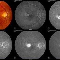
32-year-old African American male with Grade IV hypertensive retinopathy and acute renal failure. Vision OD 20/70, OS 20/25. Creatine 7.1. BP: 250/150.
Photographer: Geoffrey Pankhurst, University Hospitals, Eye Institute/Dept. Ophthalmology and Visual Sciences Case Western Reserve University Cleveland, OH
Imaging device: Topcon TRC 50x
Condition/keywords: acute renal failure, disc edema, exudate, hypertension, hypertensive retinopathy, ischemia, macular edema, macular ischemia, optic disc edema
-
 Hypertensive Retinopathy
Feb 25 2013 by Suber S. Huang, MD, MBA, FASRS
32-year-old African American male with Grade IV hypertensive retinopathy and acute renal failure. Vision OD 20/70, OS 20/25. Creatine 7.1. BP: 250/150.
Photographer: Geoffrey Pankhurst, University Hospitals, Eye Institute/Dept. Ophthalmology and Visual Sciences Case Western Reserve University Cleveland, OH
Imaging device: Topcon TRC 50x
Condition/keywords: acute renal failure, disc edema, exudate, hypertension, hypertensive retinopathy, ischemia, macular edema, macular ischemia, optic disc edema
-
 Choriodal Rupture - 003 - 6 Month Follow Up
Mar 11 2013 by Suber S. Huang, MD, MBA, FASRS
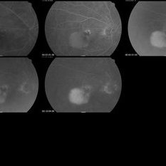
40 -year-old male sustained blunt trauma OD with orbital fracture and choroidal rupture subjacent to inferior arcade with blood, subretinal fluid, and exudate extending to fovea with CF 2 feet at presentation 9-10-12. On followup 3-11-12, vision improved to 20/400 with resolutionof hemorrhage, normal OCT, but speckling of foveal RPE and PMB consistent with damage.
Photographer: Mark Harrod
Condition/keywords: autofluorescence imaging, blunt trauma, choroidal hemorrhage, choroidal rupture, orbital fracture, retinal hemorrhage, submacular hemorrhage
-
 Choriodal Rupture 004 - Fundus Autoflurescence - 6 Month Follow Up
Mar 11 2013 by Suber S. Huang, MD, MBA, FASRS
40-year-old male sustained blunt trauma OD with orbital fracture and choroidal rupture subjacent to inferior arcade with blood, subretinal fluid, and exudate extending to fovea with CF 2 feet at presentation 9-10-12. On follow up 3-11-12, vision improved to 20/400 with resolutionof hemorrhage, normal OCT, but speckling of foveal RPE and PMB consistent with damage.
Photographer: Mark Harrod
Condition/keywords: autofluorescence imaging, blunt trauma, choroidal hemorrhage, choroidal rupture, orbital fracture, retinal hemorrhage, submacular hemorrhage
-
 Choriodal Rupture - AW 001 - Initial Presentation
Mar 11 2013 by Suber S. Huang, MD, MBA, FASRS
40-year-old male sustained blunt trauma OD with orbital fracture and choroidal rupture subjacent to inferior arcade with blood, subretinal fluid, and exudate extending to fovea with CF 2 feet at presentation 9-10-12. On followup 3-11-12, vision improved to 20/400 with resolution of hemorrhage, normal OCT, but speckling of foveal RPE and PMB consistent with damage.
Photographer: Mark Harrod
Condition/keywords: autofluorescence imaging, blunt trauma, choroidal hemorrhage, choroidal rupture, orbital fracture, retinal hemorrhage, submacular hemorrhage
-
 Choriodal Rupture - AW 002 - 6 Month F/U
Mar 11 2013 by Suber S. Huang, MD, MBA, FASRS
40-year-old male sustained blunt trauma OD with orbital fracture and choroidal rupture subjacent to inferior arcade with blood, subretinal fluid, and exudate extending to fovea with CF 2 feet at presentation 9-10-12. On follow up 3-11-12, vision improved to 20/400 with resolutionof hemorrhage, normal OCT, but speckling of foveal RPE and PMB consistent with damage.
Photographer: Mark Harrod
Condition/keywords: autofluorescence imaging, blunt trauma, choroidal hemorrhage, choroidal rupture, orbital fracture, retinal hemorrhage, submacular hemorrhage
-
 Histopathology Mouse Retina - Normal
Apr 25 2013 by Suber S. Huang, MD, MBA, FASRS
Mouse retinal structure is presented. The retina consists of seven layers, ganglion cell layer, inter plexiform layer, inner nuclear layer, outer plexiform layer, outer nuclear layer, photoreceptor layer and the retinal pigmented epithelium layer. Nuclei were stained with dapi (blue). Two kinds of photoreceptor cells; cone photoreceptors were stained with PNA (green) and rod photoreceptors were stained with anti-rhodopsin antibody (red).
Photographer: Akiko Maeda, Tadao Maeda
Imaging device: Fluorescence microscope
Condition/keywords: histopathology, retina
-
---thumb.jpg/image-square;max$300,300.ImageHandler) Age Related Macular Degeneration
Age Related Macular Degeneration
May 3 2013 by Suber S. Huang, MD, MBA, FASRS
Age related macular degeneration.
Condition/keywords: advanced geographic atrophy, atrophic scar, atrophic spot, geographic atrophy, macula lesion, pigment epithelial atrophy, red-free, window defect
-
---thumb.jpg/image-square;max$300,300.ImageHandler) Age Related Macular Degeneration - Geographic Atrophy
Age Related Macular Degeneration - Geographic Atrophy
May 3 2013 by Suber S. Huang, MD, MBA, FASRS
Geographic Atrophy.
Imaging device: Retina Diseases Imaging Analysis Reading Center
Condition/keywords: advanced geographic atrophy, atrophic scar, atrophic spot, geographic atrophy, macula lesion, pigment epithelial atrophy
-
---thumb.jpg/image-square;max$300,300.ImageHandler) Age Related Macular Degeneration - Geographic Atrophy
Age Related Macular Degeneration - Geographic Atrophy
May 3 2013 by Suber S. Huang, MD, MBA, FASRS
Geographic Atrophy.
Imaging device: Retina Diseases Imaging Reading Center
Condition/keywords: advanced geographic atrophy, atrophic scar, atrophic spot, geographic atrophy, macula lesion, pigment epithelial atrophy, red-free, window defect
-
---thumb.jpg/image-square;max$300,300.ImageHandler) Age Related Macular Degeneration - Geographic Atrophy
Age Related Macular Degeneration - Geographic Atrophy
May 3 2013 by Suber S. Huang, MD, MBA, FASRS
Geographic Atrophy.
Imaging device: Retina Diseases Imaging Analysis Reading Center
Condition/keywords: advanced geographic atrophy, atrophic scar, atrophic spot, geographic atrophy, macula lesion, pigment epithelial atrophy
-
 Age related Macular Degeneration - Classic CNV
May 3 2013 by Suber S. Huang, MD, MBA, FASRS
Classic CNV.
Imaging device: Retina Diseases Imaging Analysis Center
Condition/keywords: choroidal neovascularization (CNV), classic form
-
 Age Related Macular Degeneration
May 3 2013 by Suber S. Huang, MD, MBA, FASRS
Classic CNV.
Imaging device: Retina Diseases Imaging Analysis Reading Center
Condition/keywords: choroidal neovascularization (CNV), classic form
-
 Age Related Macular Degeneration - Occult CNV
May 3 2013 by Suber S. Huang, MD, MBA, FASRS
Age related macular degeneration - occult CNV
Imaging device: Retina Diseases Imaging Analysis Reading Center
Condition/keywords: choroidal neovascularization (CNV), choroidal neovascularization occult
-
 Age Related Macular Degeneration - Occult CNV
May 3 2013 by Suber S. Huang, MD, MBA, FASRS
Age related macular degeneration - occult CNV.
Imaging device: Retina Diseases Imaging Analysis Reading Center
Condition/keywords: choroidal neovascularization (CNV), choroidal neovascularization occult
-
 CME
CME
May 3 2013 by Suber S. Huang, MD, MBA, FASRS
CME.
Imaging device: Retina Diseases Imaging Analysis Reading Center
Condition/keywords: angiographic macular leakage, cystoid macular edema (CME), FA late phase leakage
-
 Retinoblastoma - Regressed
May 3 2013 by Suber S. Huang, MD, MBA, FASRS
24-year-old male status post radiation for retinoblastoma with secondary metastatic carcinoma.
Imaging device: Retina Diseases Imaging Analysis Reading Center
Condition/keywords: endophytic tumor growth, intraocular tumor, macular lesion, radiotherapy, retinoblastoma
-
 Primary Ocular Lymphoma
Jun 28 2012 by Suber S. Huang, MD, MBA, FASRS
Unilateral biopsy proven ocular lymphoma without systemic involvement. Father died of non-Hodgkins lymphoma. Older brother died of Hodgkin's lymphoma.
Photographer: Mark Harrod/Geoffrey Pankhurst, Case Western Reserve University/University Hospitals of Cleveland, Cleveland, OH
Imaging device: Topcon
-
 Papilledema
Sep 21 2012 by Suber S. Huang, MD, MBA, FASRS
Fundus photograph of a 24-year-old obese woman with severe papilledema secondary to idiopathic intracranial hypertension.
Condition/keywords: dilated tortuous vessels, exudate, idiopathic intracranial hypertension, Paton's lines, peripapillary hemorrhage, pseudotumor cerebri
-
 Optic Disc Drusen
Sep 21 2012 by Suber S. Huang, MD, MBA, FASRS
Fundus photograph of a 50-year-old woman with optic disc drusen complicated by anterior ischemic optic neuropathy
Condition/keywords: optic disc drusen
-
 Optic Disc Drusen
Sep 21 2012 by Suber S. Huang, MD, MBA, FASRS
Optic disc drusen demonstrating autofluorescence
-
 Chocolate Eye
Oct 8 2012 by Suber S. Huang, MD, MBA, FASRS
Useful model for anatomy, patient education
Photographer: Daniel Huang
Condition/keywords: best Xmas present ever, model eye, saggital section of milk chocholate eye
-
 STAR syndrome
Dec 14 2012 by Suber S. Huang, MD, MBA, FASRS
Fundus photograph of a 32-year-old woman with STAR syndrome (Syndactyly, Telecanthus, Anogenital and Renal Malformations) and good vision; SD-OCT with normal findings; Also uploaded are RetCam images of patient's 5 year old daughter with STAR syndrome
Photographer: Mark Herrod
-
---thumb.jpg/image-square;max$300,300.ImageHandler) STAR Syndrome
STAR Syndrome
Dec 14 2012 by Suber S. Huang, MD, MBA, FASRS
Fundus photograph of a 32-year-old woman with STAR syndrome (Syndactyly, Telecanthus, Anogenital and Renal Malformations) and good vision; SD-OCT with normal findings; Also uploaded are RetCam images of patient's 5 year old daughter with STAR syndrome
Photographer: Mark Herrod
A project from the American Society of Retina Specialists