-
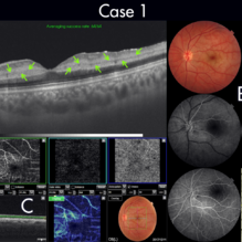 Figure-1 Paracentral Acute Middle Maculopathy (PAMM)
Dec 21 2018 by Fawwaz F Al Mamoori, MD, Medical Retina Consultant
25-year-old male patient medically free, had sudden deterioration in his left eye vision. Visual acuity on presentation was counting fingers at 3 meter distance. Marked Relative Pupillary Afferent Defect (RAPD) was detected and fundoscopic exam showed abnormal foveal reflex. SS OCT B scan: showed a hypereflectivity of the inner plexiform layer (IPL), inner nuclear layer (INL) and OPL layer (fig 1, A).FA images were normal (fig 1, B). Angiography shows remarkable perifoveal capillary drop out within middle retinal layer correlating with perfusion density map which reveals significant decrease in capillary density at the same level (Fig 1, C). Enface ads more proof to PAMM by delineating ischemic distribution in a fern like pattern of hyper reflective areas within DCP (fig1, D).
Photographer: Dr.Fawwaz Al Mamoori (Al Mamoori Eye Clinic)
Imaging device: Triton Swept Source OCT (TOPCON)
Condition/keywords: optical coherence tomography (OCT), paracentral acute middle maculopathy
-
 Resolving of Refractory Subretinal Fluid Post Faricimab Injection
Nov 7 2022 by Fawwaz F Al Mamoori, MD, Medical Retina Consultant
A case of Rt eye wet-AMD treated over 2 years with a Q8weeks aflibercebt injection with refractory SRF less than 200 um that shifted recently to Faricimab.1 month post Faricimab OCT showed a complete drying out of refractory subretinal Fluid (SRF)
Photographer: Mohammed Rabaa
Imaging device: Swept Source OCT (TRITON) Topcon
Condition/keywords: subretinal fluid
-
 Non invasive multimodal imaging for differentiating unilateral pseudo swelling buried optic disc drusen from true optic disc swelling
Feb 7 2024 by Fawwaz F Al Mamoori, MD, Medical Retina Consultant
27-year-old male, medically free, routine fundus examination showed left optic dic swelling, BCVA =1.0(OU), color vision, and contrast sensitivity were normal with no RAPD (OU). SS-OCT of the left optic disc showed a hyporeflective mass. Enface OCT shadogram showed peripapillary ovoid structures (drusen).FAF: showed drusenoid autofluorescence in the superonasal part only. Orbital MRI with contrast was requested to exclude any optic nerve tumor and it was normal.
Photographer: Hana.S.Owais
Imaging device: TRITON(OCT) Topcon
Condition/keywords: multimodal imaging, optic disc drusen, optic disc swelling
-
 Multimodal Imaging for Differentiating Unilateral Pseudo Optic Disc Swelling(Buried Drusen) From True Optic Disc Swelling
Feb 7 2024 by Fawwaz F Al Mamoori, MD, Medical Retina Consultant
A 27-year-old male patient, medically free, presented with unilateral left optic disc swelling. BCVA=1.0(OU), color vision, and contrast sensitivity were normal (OU) with no RAPD in the left eye. SS-OCT: showed left optic disc elevation with hyporeflective mass lesion (Fig-1 B). Enface OCT: showed left peripapillary hyperreflective ovoid mass lesions(Fig-2 D, Fig-3 F), FAF: showed left superonasal hyperautofluorescent drusenoid lesions. Orbital MRI with contrast was requested to exclude any optic nerve compressive lesions like (tumors: like mengioma or inflammatory lesions like granuloma (sarcoidosis). the result of orbital MRI was normal.
Photographer: Hana.S.Owais
Imaging device: TRITON(TOPCON,Swept Source OCT)
Condition/keywords: fundus autofluorescence (FAF), multimodal imaging, OCT EN FACE, optic disc drusen, optic disc edema
-
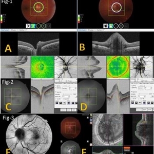 Multimodal Imaging for Differentiating Unilateral Pseudo Optic Disc Swelling(Buried Drusen) From True Optic Disc Swelling
Feb 7 2024 by Fawwaz F Al Mamoori, MD, Medical Retina Consultant
27-year-old male, medically free, presented with left unilateral optic disc swelling. BCVA=1.0(OU), color vision, and contrast sensitivity were normal (OU)with no RAPD in the left eye. Swept Source OCT: showed elevated left optic disc with hyporeflective mass (Fig-1 B). Enface OCT: Showed left peripapillary multiple ovoid mass lesions(drusen) (Fig-2 d, Fig3 F). FAF: of the left eye showed superonasal hyper autofluorescent drusenoid lesions)(Fig3 E). Orbital MRI with contrast was requested to exclude any compressive lesions like tumors(menigioma)or inflammatory lesions like granuloma(sarcoid granuloma). orbital MRI result was normal.
Photographer: Hana.S.Owais
Imaging device: TRITON(TOPCON,Swept Source OCT)
Condition/keywords: fundus autofluorescence (FAF), multimodal imaging, OCT EN FACE, optic disc drusen, optic disc edema, swept source
A project from the American Society of Retina Specialists