File number: 28275
Comments
-
 Hosam Attia, MD (June 18 2018)
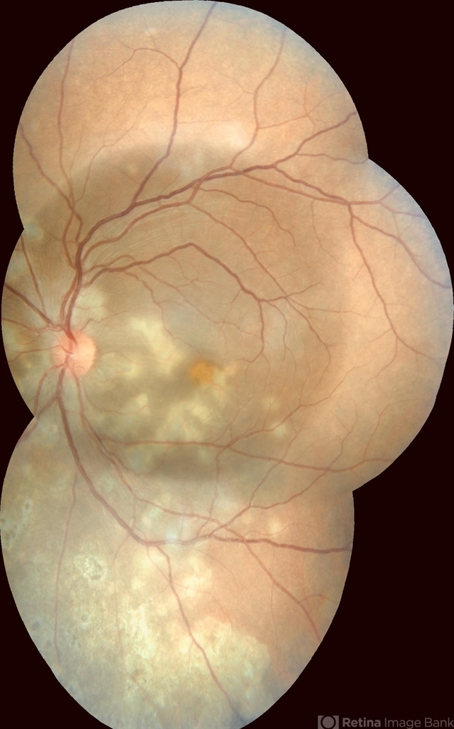
They appear to be at different stages of healing though, with the peripheral inferior ones, appear inactive, older & atrophic.
I am sure the darker, perfect circle in the middle, is just an artifact due to different exposure and not an exudative RD, as it is too perfectly round (corresponding with central color photo frame, with the active choroidal lesions mostly below.
Could this be Serpiginous Choroiditis ??
Just out of curiosity, did you obtain FA +/- ICG and OCT through some of these lesions, i wonder what it looks like ?
Thanks for sharing
Sign in to comment.
 Initializing download.
-
 By Purva Patwari
Patwari Retina Clinic - Uploaded on Jun 13, 2018.
- Last modified by Caroline Bozell on Jun 14, 2018.
- Rating
- Appears in
- Patwari Retina Clinic
- Condition/keywords
- choroiditis, disseminated choroiditis
- Photographer
- Dr Purva Patwari, Patwari Retina Center, Ahmedabad, Gujarat , India
- Imaging device
-
Fundus camera
Zeiss Visucam 500 - Description
- A 25-year-old male patient presented with defective vision noticed since last 2 weeks. No significant past history. Anterior segment was normal. Vision RE 6/6, LE 6/60. Other eye was normal on examination.


