-
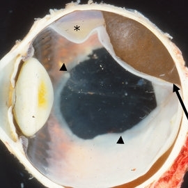 Pigmented Choroidal Melanoma
May 18 2020 by McGill University Health Centre
Choroidal melanoma is often asymptomatic and diagnosis is incidental. The tumors may grow beneath the retina, or may break through the Bruch membrane and disrupt the retina. Tumors breaking through the Bruch membrane and disrupting the retina have a characteristic “mushroom” shape. This enucleation specimen shows a pigmented, dome-shaped choroidal melanoma (arrow) with serous retinal detachment (*). Note the deeply pigmented area in the retina, which corresponds to hyperplastic pigment epithelium proliferation (arrowheads). In general, this proliferation is congenital and not related to the tumor.
Condition/keywords: pigment
-
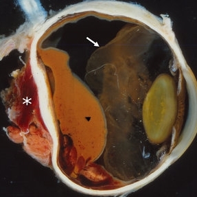
 Choroidal Melanoma
May 18 2020 by McGill University Health Centre
Choroidal melanoma is often asymptomatic and diagnosis is incidental. The tumors may grow beneath the retina, or may break through the Bruch membrane and disrupt the retina. Tumors breaking through the Bruch membrane and disrupting the retina have a characteristic “mushroom” shape. This enucleation specimen shows a pigmented dome-shaped choroidal melanoma (arrow). The cataractous lens is dislocated (*) and the retina is folded (•).
Condition/keywords: enucleation
-
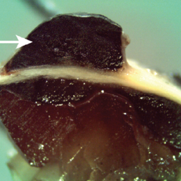 Choroidal Melanoma with Extraocular Extension
May 18 2020 by McGill University Health Centre
Choroidal melanoma is often asymptomatic and diagnosis is incidental. The tumors may grow beneath the retina, or may break through the Bruch membrane and disrupt the retina. Tumors breaking through the Bruch membrane and disrupting the retina have a characteristic “mushroom” shape. This enucleation specimen demonstrates a section of a choroidal melanoma showing an intraocular tumor with an extraocular extension (arrow).
Condition/keywords: extraocular extension
-
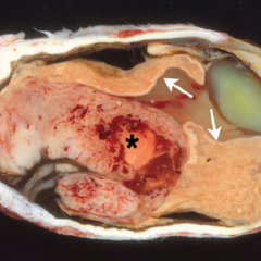
 Choroidal Melanoma with Extraocular Extension
May 18 2020 by McGill University Health Centre
Choroidal melanoma is often asymptomatic and diagnosis is incidental. The tumors may grow beneath the retina, or may break through the Bruch membrane and disrupt the retina. Tumors breaking through the Bruch membrane and disrupting the retina have a characteristic “mushroom” shape. The diffuse melanoma seen in this enucleation specimen is infiltrating the choroid. Extensive areas of necrosis and hemorrhage are present (arrowhead). The extraocular involvement is large and surrounded by hemorrhage (*). The retina is folded (arrow).
Condition/keywords: extraocular extension
-
 Amelanotic Choroidal Melanoma
May 18 2020 by McGill University Health Centre
The enucleation specimen in (A) shows an amelanotic, mushroom-shaped tumor arising from the choroid. Microhemorrhages are present within the tumor and also surround the tumor base (arrow). True retinal detachment is present (arrowhead). The subretinal fluid is mixed: clear (1), hemorrhagic (2), and fibrinoid (3).
Condition/keywords: enucleation, mushroom-shaped
-
 Amelanotic Choroidal Melanoma
May 18 2020 by McGill University Health Centre
The enucleation image shows a large amelanotic tumor with large areas of hemorrhage and necrosis. Note the several dilated blood vessels and an adjacent retinal detachment with lipofuscin pigment on its surface (arrow).
Condition/keywords: amelanotic melanoma, enucleation, mushroom-shaped
-
 Amelanotic Mushroom-Shaped Choroidal Melanoma
May 18 2020 by McGill University Health Centre
This enucleation specimen demonstrates an amelanotic, mushroom-shaped, slightly hemorrhagic tumor near the optic nerve (arrow). True retinal detachment is present, and the retina is folded (arrowhead). The subretinal fluid is hazy (*).
Condition/keywords: amelanotic melanoma, enucleation, mushroom-shaped
-
 Amelanotic Mushroom-Shaped Choroidal Melanoma
May 18 2020 by McGill University Health Centre
The enucleation specimen in (B) shows an amelanotic, mushroom-shaped, slightly hemorrhagic tumor near the optic nerve (arrow). The shape is due to infiltration of the retina by a rupture of the Bruch membrane. A retinal detachment artifact is present.
Condition/keywords: enucleation, mushroom-shaped
-
 Mushroom-shaped Choroidal Melanoma
May 18 2020 by McGill University Health Centre
The enucleation specimen in (A) shows a diffuse melanoma infiltrating the choroid and ciliary body. In the center, a large area of necrosis and hemorrhage is present (*) and the retina is infiltrated (arrows). Note hypopyon in the anterior chamber and the cataractous lens.
Condition/keywords: enucleation, mushroom-shaped
A project from the American Society of Retina Specialists