-
---thumb.jpg/image-square;max$300,300.ImageHandler) Myopic Giant Tear
Myopic Giant Tear
Mar 13 2014 by Marcelo Zas, MD PhD
The image show a myopic giant tear with irregular edges.
Photographer: Marcelo Zas MD PhD
Condition/keywords: giant retinal tear, myopia
-
 PVR With Multiple Breaks
Mar 13 2014 by Marcelo Zas, MD PhD
The image show a PVR case with PFCL in the posterior pole, diathermy is used before the retinectomy.
Photographer: Marcelo Zas MD PhD
Condition/keywords: proliferative vitreoretinopathy (PVR)
-
---thumb.jpg/image-square;max$300,300.ImageHandler) Retinectomy With Diathermy in a Giant Tear
Retinectomy With Diathermy in a Giant Tear
Mar 13 2014 by Marcelo Zas, MD PhD
The image show a giant tear in a myopic patient. We use diathermy to avoid intraop bleeding.
Photographer: Marcelo Zas MD PhD
Condition/keywords: giant retinal tear, myopia
-
 Choroidal Detachment
Oct 1 2021 by Marcelo Zas, MD PhD
Right eye from a 65-year-old patient with a choroidal detachment post trabeculectomy.
Photographer: Zas Marcelo MD, PhD
Imaging device: Optos California
Condition/keywords: choroidal detachment, post-trabeculectomy
-
 Intraretinal cysts
Nov 15 2021 by Marcelo Zas, MD PhD
Left eye from a young patient with a chronic rhegmatogenous retinal detachment presenting intraretinal cysts.
Photographer: Zas Marcelo MD PhD
Condition/keywords: chronic retinal detachment, intraretinal cyst
-
 Intraretinal cysts
Nov 15 2021 by Marcelo Zas, MD PhD
Left eye from a young patient with a chronic rhegmatogenous retinal detachment presenting intraretinal cysts.
Photographer: Zas Marcelo MD PhD
Condition/keywords: chronic retinal detachment, intraretinal cyst
-
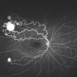
 Von Hippel Lindau with retinal capillary hemangioma
Nov 2 2023 by Marcelo Zas, MD PhD
30-year-old female patient diagnosed with Syndrome VHL (Von Hippel Lindau). Stage II. In the first wide-field retinography of the right eye we can observe the exophytic retinal hemangiomas, rounded, slightly delimited, located in the peripheral retina in the upper and lower temporal quadrants and due to the exudation produced by them, hard exudates are observed in the star hemisphere, affecting the macula.
Photographer: Mariano Cotic MD
Imaging device: Silverstone SS OCT Optos
Condition/keywords: abnormal retinal vessel
-
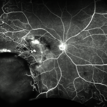
 Von Hippel Lindau With Retinal Capillary Hemangioma
Nov 2 2023 by Marcelo Zas, MD PhD
This wide-field AGF image shows the vascular tumors with their corresponding afferent and efferent vessels.
Photographer: Mariano Cotic MD
Imaging device: Silverstone SS OCT Optos
Condition/keywords: tumor
-
 Best Disease
Apr 24 2024 by Marcelo Zas, MD PhD
Best vitelliform macular dystrophy (BVMD) or Best disease. Is the most common autosomal dominant macular dystrophy. It involves the retinal pigment epithelium (RPE), and leads to a characteristic bilateral yellow “egg-yolk” appearance of the macula as you can see in this image. Essentially, BVMD is considered to have 6 clinical stages: Previtelliform, Vitelliform, Pseudohypopyon, Vitelleruptive, Atrophic and Choroidal neovascularization. As the disease progresses, patients may experience a slow, bilateral decrease in visual acuity, central scotoma, or metamorphopsia. With secondary CNV, visual decline can be rapid, however.
Photographer: Luciano Scorsetti MD
Condition/keywords: Macular Dystrophy
-
 Proliferative Vitreoretinopathy
Jun 9 2024 by Marcelo Zas, MD PhD
We present a case of a 20-year-old patient who underwent surgery for congenital cataract when he was born and 20 years after he developed a retinal detachment with proliferative vitreoretinopathy. Proliferative vitreoretinopathy (PVR), a major complication of rhegmatogenous retinal detachment (RRD), is an abnormal process whereby proliferative, contractile cellular membranes form in the vitreous and on both sides of the retina, resulting in tractional retinal detachment with fixed retinal folds. PVR arises in an estimated 5-10% of RRD cases, and therefore represents a major complication of retinal detachment. The best treatment of PVR is its prevention. Clinical factors associated with increased risk of PVR include: • Chronic RRD • 2 o more horseshoe retinal tears and RRD exposing three-disc diameters or more of RPE • RD associated with giant retinal • RD associated with choroidal detachment • Ocular Trauma • RRD associated with vitreous hemorrhage • Aphakia and RRD • Failure of previous surgery or multiple retinal surgeries • Aggressive retinitis, etc.
Photographer: Luciano Scorsetti MD
Condition/keywords: proliferative vitreoretinopathy (PVR)
-
 Retinal Vasoproliferative Tumor
Jun 24 2025 by Marcelo Zas, MD PhD
We present a case of a 33-year-old male patient, who presented with decreased visual acuity in his right eye with 20/80, presenting a primary retinal vasoproliferative tumor in the lower temporal quadrant. The tumor is associated with serous retinal detachment, hard exudation, neovascularization and telangiectasias. Lipid exudates extend toward the macula, indicating macular involvement, which may contribute to decreased visual acuity. Oi was normal with 20/20 of BCVA. The patient was treated initially with IV anti-VEGF therapy and cryotherapy.
Photographer: Marcelo Zas MD PhD
Condition/keywords: RETINAL VASOPROLIFERATIVE TUMOR
-
 Fluorescein Angiography (FA) of a Primary Retinal Vasoproliferative Tumor
Jun 29 2025 by Marcelo Zas, MD PhD
We present a case of a 33-year-old male patient, who presented with decreased visual acuity in his right eye with 20/80, presenting a primary retinal vasoproliferative tumor in the lower temporal quadrant. The fluorescein angiography findings are: 1. Early hyperfluorescence due to its rich intrinsic vascularity and often has dilated feeding arterioles and draining venules. 2. Marked progressive leakage from the tumor vessels. 3. The late leakage often obscures fine vascular details in the late phase and corresponds to exudation and macular edema seen clinically. 4. Staining of surrounding exudates, RPE disturbances and gliosis. 5. In our case also a marked peripheral capillary closure in the areas adjacent to the tumor and in other quadrants as well.
Photographer: Marcelo Zas MD PhD
Condition/keywords: RETINAL VASOPROLIFERATIVE TUMOR
A project from the American Society of Retina Specialists