 Initializing download.
-
 By RUSHIK PATEL
NETRALAYA SUPER SPECIALITY EYE HOSPITAL - Uploaded on Jun 21, 2022.
- Last modified by Joshua Friedman on Jun 22, 2022.
- Rating
- Appears in
- 21-Jun-2022
- Condition/keywords
- tractional retinal detachment, proliferative diabetic retinopathy (PDR)
- Photographer
- Dr Rushik Patel, Netralaya super-speciality eye hospital
- Imaging device
- Optical coherence tomography system
- Description
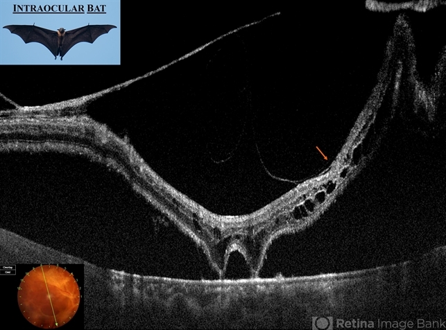
- The swept-source optical coherence tomography of right eye of 45 year old diabetic female having proliferative diabetic retinopathy (PDR) with long standing tractional retinal detachment showed pre-retinal fibrovascular membrane causing tractional neurosensory retinal (NSR) detachment involving macula (orange arrow) except 2 focal attachments with underlying retinal pigment epithelium (RPE). There were chronic cystoid changes in the detached NSR. The tractional elevation of NSR surrounding 2 focal attachments with RPE gives an appearance resembling "BAT".


---thumb.jpg/image-square;max$79,0.ImageHandler "PDR Neovacularization of the Disc")


---thumb.JPG/image-square;max$79,0.ImageHandler "Neovascularisation Disc")
