-
 Acute Posterior Multifocal Placoid Pigment Epitheliopathy (APMPPE)
Oct 8 2020 by Mihir Trivedi
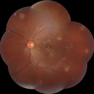
Fundus photograph of a 21-year-old male having complaints of blurring of vision. Had a viral prodrome 2 weeks before the onset of symptoms.
Imaging device: DRI OCT-1 Triton (plus) TOPCON
Condition/keywords: acute multifocal placoid pigment epitheliopathy (AMPPE)
-
 Central Areolar Choroidal Dystrophy
Oct 30 2020 by Mihir Trivedi
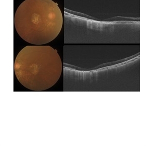
Fundus photo of a 43-year-old female with gradual onset diminution of vision in both eyes since 2-3 years. BCVA in OU was 3/60. She was diagnosed to have central areolar choroidal dystrophy(CACD). Central areolar choroidal dystrophy (CACD) is a rare inherited disease, which causes progressive profound loss of vision in patients during their fourth decade. It is characterized by atrophy of retinal pigment epithelium, photoreceptors and choriocapillaris. IT is a progressive macular dystrophy characterized by subtle, mottled depigmentation in the posterior pole in the early stages. The depigmentation area gradually enlarges until an oval or round surface of atrophy of the retinal pigmentary epithelium and choriocapillaris is formed. Drusen or flecks are absent in a typical presentation.
Photographer: Mr Ganesh Naidu
Imaging device: TOPCON DRI Triton
Condition/keywords: central areolar choroidal dystrophy (CACD)
-
 Unilateral Macular Coloboma
Jul 29 2021 by Mihir Trivedi
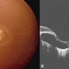
Fundus examination of a 35-year-old man with focal areas of altered retinal pigment epithelium and subretinal yellowish lesion in the foveal area in the right eye. Left eye showed a punched out circumscribed lesion in the center of the macula with thin foveal roof suggestive probably of the internal limiting membrane. Macular coloboma is characterized by a sharply defined, oval or rounded, usually unilateral, atrophic lesions of varying size presenting rudimentary or absent retina, choroid and sclera located at the macula leading to decreased vision in the central area of the fundus. It can be associated with retinal dystrophy in the fellow eye, as was the case in our patient.
Photographer: Priyanshi Kambodi, RNC Eye Hospital, Valsad
Condition/keywords: macular coloboma
A project from the American Society of Retina Specialists