 Initializing download.
-
 By Aditya Verma, MBBS, MS, Post fellow
Medical Research Foundation, Sankara Nethralaya
Co-author(s): Vikas Khetan - Uploaded on Jun 5, 2021.
- Last modified by Caroline Bozell on Jun 11, 2021.
- Rating
- Appears in
- 5-Jun-2021
- Condition/keywords
- macular edema, macular exudates, hard exudates
- Photographer
- Aditya Verma
- Imaging device
-
Scanning laser ophthalmoscope
Heidelberg Spectralis - Description
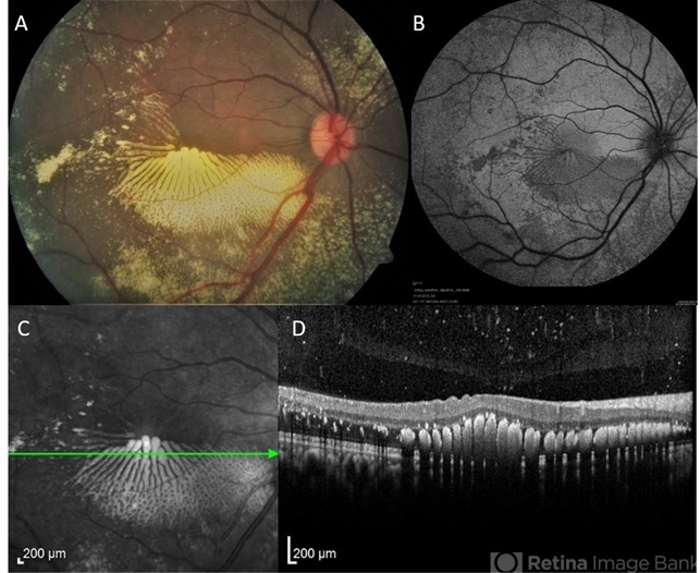
- Multimodal imaging of a 36-year-old male with peripheral vasoproliferative tumor. Posterior pole showed exquisite pattern of hard exudate accumulation in petaloid pattern (A), fundus autofluorescence (B) and infrared image (C) portrayed the precise pattern of hard exudate distribution; Optical coherence tomography scan (D) showed a uniformly distributed parallel clumps of exudates in the outer plexiform layer.


---thumb.jpg/image-square;max$79,0.ImageHandler "Leber's Stellate Maculopathy")