-
 Macular Edema
Jun 5 2021 by Aditya Verma, MBBS, MS, Post fellow
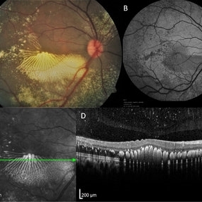
Multimodal imaging of a 36-year-old male with peripheral vasoproliferative tumor. Posterior pole showed exquisite pattern of hard exudate accumulation in petaloid pattern (A), fundus autofluorescence (B) and infrared image (C) portrayed the precise pattern of hard exudate distribution; Optical coherence tomography scan (D) showed a uniformly distributed parallel clumps of exudates in the outer plexiform layer.
Photographer: Aditya Verma
Imaging device: Heidelberg Spectralis
Condition/keywords: hard exudates, macular edema, macular exudates
A project from the American Society of Retina Specialists