 Initializing download.
-
 By Priya Rasipuram Chandrasekaran, MBBS, DO, DNB, FRCS
Lotus eye hospital
Co-author(s): Lotus eye hospital, Salem - Uploaded on May 4, 2021.
- Last modified by Caroline Bozell on May 4, 2021.
- Rating
- Appears in
- Miscellaneous
- Condition/keywords
- chorioretinitis sclopetaria
- Imaging device
- Fundus camera
- Description
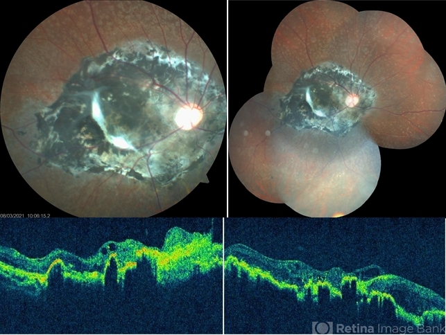
- This fundus photo and montage shows pigmentary changes with fibroglial proliferation of the disc and macula in a 36-year-old male following injury with an iron chain. This is usually following a high velocity non-penetrating missile or blast injury categorized as coup injury and can be both direct or indirect. The layers affected are the highly inelastic Bruch’s membrane with choriocapillaris and retinal pigment epithelium in contrast to the highly elastic retina and sclera. The high impact injury causes full thickness defect in the retina, Bruch’s membrane and choroid leading to retraction of the retina and choroid, leaving the intact bare sclera behind. Pathology included defects in the Bruch’s membrane and choroid, and extensive photoreceptor loss with hyperplasia of retinal pigment epithelium. Over the weeks, loose fibrous tissue gets replaced by dense connective tissue leading to scarring between retina and choroid as seen in our patient. The background shows fundus albipunctatus.


