 Initializing download.
-
 By Deependra Vikram Singh, MD FASRS
Eye-Q Superspecialty Eye Hospitals
Co-author(s): Raja Rami Reddy, Neoretina Institute, Hyderabad, INDIA - Uploaded on Jul 7, 2020.
- Last modified by Caroline Bozell on Jul 7, 2020.
- Rating
- Appears in
- Miscellaneous
- Condition/keywords
- encapsulated intraocular foreign body
- Photographer
- Deependra Vikram Singh, Eye-Q Hospitals, Gurugram, INDIA
- Imaging device
-
Fundus camera
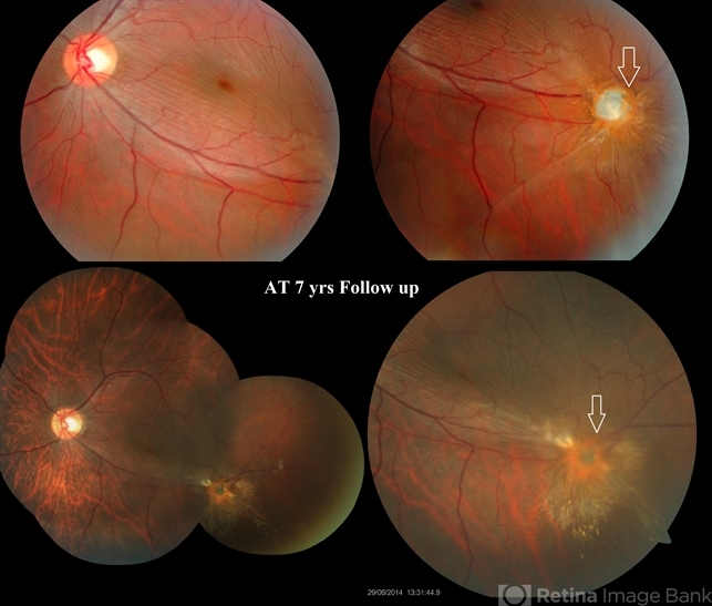
Kowa and Zeiss - Description
- 25-yr-old male presented to our retina clinic in 2007 with history of Hammer and chisel injury to left eye 2 months back. On examination BCVA in left eye was 20/20. Slit-lamp examination revealed iris hole and fundus examination showed an encapsulated metallic intraocular foreign body (IOFB) close to inferior arcade in left eye. Patient was advised Vitreous surgery with IOFB removal. Patient, however did not turn up for Surgery and revisited our clinic after seven years in 2014. On examination his BCVA was 20/20 in left eye and IOFB has reduced in size with brown siderotic deposits seen over IOFB capsule. Examination revealed posterior sub-capsular cataract but no siderotic changes with intraocular pressure (IOP) also being recorded as normal. In view of good visual acuity and no siderotic changes, he was advised regular follow up and ERG. Since most IOFBs would get timely removed by vitreous surgery, this Image capturing the natural course of a metallic IOFB is rare.


