 Initializing download.
-
 By Olivia Rainey
Retina Specialists of Michigan
Co-author(s): Joseph Boss, MD - Uploaded on Jun 15, 2020.
- Last modified by Caroline Bozell on Jun 16, 2020.
- Rating
- Appears in
- Miscellaneous
- Condition/keywords
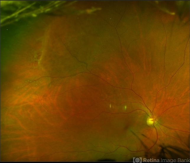
- bullous retinoschisis, Optos, outer layer breaks, outer layer hole, subretinal fluid, pseudocolor, ultra-wide field imaging, superior retina
- Photographer
- Olivia Rainey, OCT-C, COA
- Imaging device
-
Fundus camera
Optos California - Description
- Ultra-widefield pseudocolor fundus photograph of a 56-year-old female with bullous retinoschisis with outer retinal holes affecting her right eye. The physician noted superotemporal retinoschisis in her monoculcar functioning eye. There was no demarcation line and no inner or outer layer breaks at her first appointment in February of 2020. On 6/15/20 she had a new onset outer holes and SRF tracking inferiorly. The physician recommended observation, however if this continues to progress we have discussed indications for barrier laser.


---thumb.JPG/image-square;max$79,0.ImageHandler "Central Serous Retinopathy with Fibrin - Mid-phase FA")

---thumb.JPG/image-square;max$79,0.ImageHandler "Central Serous Retinopathy with Fibrin")