 Initializing download.
-
 By McGill University Health Centre
The MUHC-McGill University
Co-author(s): Sabrina Bergeron, P. Zoroquiain, E. Esposito, S. Corredor Casas, P. Logan, A. N. Odashiro, Miguel N. Burnier, Paulina García de Alba Graue, McGill University Health Center-McGill University Ocular Pathology & Translational Research Laboratory - Uploaded on May 18, 2020.
- Last modified by Caroline Bozell on May 19, 2020.
- Rating
- Appears in
- Choroidal Melanoma
- Condition/keywords
- pigment
- Description
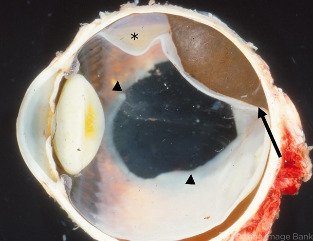
- Choroidal melanoma is often asymptomatic and diagnosis is incidental. The tumors may grow beneath the retina, or may break through the Bruch membrane and disrupt the retina. Tumors breaking through the Bruch membrane and disrupting the retina have a characteristic “mushroom” shape. This enucleation specimen shows a pigmented, dome-shaped choroidal melanoma (arrow) with serous retinal detachment (*). Note the deeply pigmented area in the retina, which corresponds to hyperplastic pigment epithelium proliferation (arrowheads). In general, this proliferation is congenital and not related to the tumor.

---thumb.jpg/image-square;max$79,0.ImageHandler "Subretinal & Epiretinal Pigment")


")


