 Initializing download.
-
 By McGill University Health Centre
The MUHC-McGill University
Co-author(s): Sabrina Bergeron, P. Zoroquiain, E. Esposito, S. Corredor Casas, P. Logan, A. N. Odashiro, Miguel N. Burnier, Paulina García de Alba Graue, McGill University Health Center-McGill University Ocular Pathology & Translational Research Laboratory - Uploaded on May 18, 2020.
- Last modified by Caroline Bozell on May 19, 2020.
- Rating
- Appears in
- Enucleated Eye
- Condition/keywords
- cross-section, enucleation, aqueous chamber, posterior chamber intraocular lens (PCIOL), vitreous chamber
- Description
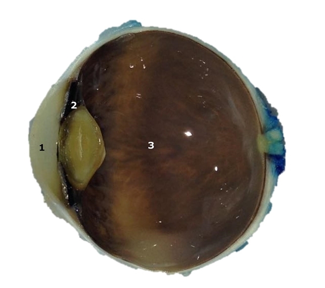
- The eye is an organ with 3 layers (from the outermost to the innermost): fibrous layer (sclera and cornea), uveal tract (iris, ciliary body, and choroid), and retina. The lens divides the eye into the aqueous chamber (filled with aqueous humor) and vitreous chamber (filled with vitreous humor). The iris divides the aqueous chamber into anterior and posterior chambers. This image illustrates the aqueous chamber (1), posterior chamber (2), and vitreous chamber (3).


---thumb.jpg/image-square;max$79,0.ImageHandler "Secondary Posterior Chamber Intraocular Lens (PCIOL)")


