-
 Right Enucleated Eye
May 18 2020 by McGill University Health Centre
Anterior view of a right enucleated eye showing the cornea, which is void in shape with a longer horizontal length (dotted lines) and the insertion points of the 4 rectus muscles (arrowheads).
Condition/keywords: cornea, enucleation
-
 Right Enucleated Eye
May 18 2020 by McGill University Health Centre
Anterior view of a right enucleated eye showing that the distance between the insertion of the rectus muscle and the limbus increases in a clockwise fashion (nasal, inferior, temporal, superior).
Condition/keywords: enucleation, insertion of the limbus, insertion of the rectus muscle
-
 Right Enucleated Eye
May 18 2020 by McGill University Health Centre
Posterior view of an enucleated eye showing the ciliary arteries marking the horizontal plane and the thick muscular insertion of the inferior oblique muscle, indicating the inferotemporal quadrant.
Condition/keywords: ciliary, enucleation, inferotemporal quadrant, insertion of the inferior oblique muscle
-
 Cross-Section of Enucleated Specimen
May 18 2020 by McGill University Health Centre
The eye is an organ with 3 layers (from the outermost to the innermost): fibrous layer (sclera and cornea), uveal tract (iris, ciliary body, and choroid), and retina. The lens divides the eye into the aqueous chamber (filled with aqueous humor) and vitreous chamber (filled with vitreous humor). The iris divides the aqueous chamber into anterior and posterior chambers. This image illustrates the aqueous chamber (1), posterior chamber (2), and vitreous chamber (3).
Condition/keywords: aqueous chamber, cross-section, enucleation, posterior chamber intraocular lens (PCIOL), vitreous chamber
-
 Enucleated Eye Showing Choroidal Melanoma
May 18 2020 by McGill University Health Centre
This enucleation specimen shows an aphakic eye with a large, solid choroidal tumor. The tumor is heavily pigmented; it shows different shades in some areas. The tumor reaches the ciliary body.
Condition/keywords: aphakic eye, choroidal tumor, enucleation
-
 Enucleated Eye with Cataractous Lens
May 18 2020 by McGill University Health Centre
The cornea is transparent and thin. The lens is cataractous. The ora Serrata (arrow) demarcates a transition zone in the uveal tract between the pars plana of the ciliary body and the retina.
Condition/keywords: cataract, enucleation
-
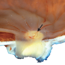
 Magnification of Enucleated Eye and Iridocorneal Angle
May 18 2020 by McGill University Health Centre
Image showing a higher magnification of the iridocorneal angle of the same specimen. The anterior chamber (1) is separated from the posterior chamber (2) by the iris. The zonulae (3) hold the lens in place. Note the pars plicata of the ciliary body (arrow).
Condition/keywords: anterior chamber, enucleation, iridocorneal angle, pars plicata, posterior chamber intraocular lens (PCIOL), zonulae
-
 Magnification of the Eye Wall from an Enucleated Eye
May 18 2020 by McGill University Health Centre
Magnification of the eye wall showing the neurosensory retina (1), the retinal pigment epithelium (arrow), a thin layer overlying the choroid (2), and the sclera (3).
Condition/keywords: choroid, enucleation, neurosensory retina, retinal pigment epithelium, sclera
-
 Enucleated Cataractous Lens Seen Through the Pupil
May 18 2020 by McGill University Health Centre
The anterior view of enucleated eye shows a cataractous lens seen through the pupil.
Condition/keywords: cataract, enucleation
-
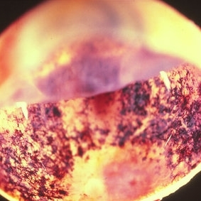
 Posterior View of Enucleated Eye with Extraocular Extension of an Intraocular Tumor
May 18 2020 by McGill University Health Centre
Posterior view of enucleated eye showing the extraocular extension of an intraocular tumor and extensive areas of hemorrhage, including a subdural hemorrhage.
Condition/keywords: enucleation, extraocular extension, intraocular tumor
-
 Enucleated Eye: Phthisis Bulbi
May 18 2020 by McGill University Health Centre
The enucleation specimen shows an atrophic eye (< 16 mm in its largest dimension) with complete disorganization of the posterior structures.
Condition/keywords: enucleation, phthisis bulbi
-
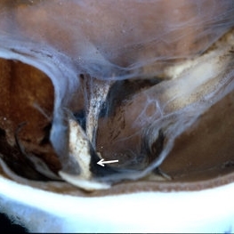 Magnification of Enucleated Eye: Phthisis Bulbi
May 18 2020 by McGill University Health Centre
Higher magnification of enucleated eye with phthisis bulbi revealing bone spicules (arrow) corresponding to ossification due to metaplastic changes of the retinal pigmented epithelium (RPE) cells toward osteoblasts.
Condition/keywords: phthisis bulbi
-
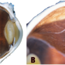 Enucleated Specimen with Pseudo-Retinal Detachment
May 18 2020 by McGill University Health Centre
In (A), a pupil–optic view of an enucleation specimen shows pseudo–retinal detachment. Note the absence of fluid beneath the retina (arrow). In (B), high magnification of a pseudo–choroidal detachment shows the bare sclera separate from the choroid and retinal pigmented epithelium.
Condition/keywords: choroidal detachment, enucleation
-
 Iridocorneal Endothelial Syndrome Enucleated Eye
May 18 2020 by McGill University Health Centre
This case of iridocorneal endothelial syndrome shows cupping of the optic nerve consistent with glaucoma (arrow).
Condition/keywords: enucleation, iridocorneal endothelial syndrome, optic nerve cupping
-
 Enucleated Eye with Subretinal Hematoma
May 18 2020 by McGill University Health Centre
This enucleation specimen shows a subretinal hematoma (arrow). In addition, a diffuse, flat retinal detachment artifact is present. The lens is cataractous.
Condition/keywords: cataract, subretinal hemorrhage
-
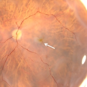 Enucleated Eye with Macular Edema
May 18 2020 by McGill University Health Centre
In an ophthalmoscopic-like view of an enucleation specimen shows macular edema. Note the folds surrounding the foveal area (arrow).
Condition/keywords: enucleation, macular edema
-
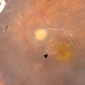 Enucleated Eye with Macular Edema
May 18 2020 by McGill University Health Centre
Image showing a specimen with irregular pigmentation of the macular area, which suggests age-related macular degeneration. The white, rounded shapes (arrowhead) may correspond to laser treatment. Note the air inside the retinal vessels.
Condition/keywords: age-related macular degeneration (AMD), macular edema
-
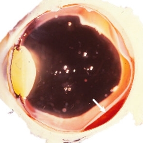
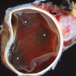 Enucleated Eye with Subretinal Hematoma
May 18 2020 by McGill University Health Centre
This enucleation specimen shows a diffuse and extended choroidal hemorrhage (*). Note the detached retina and extraocular hemorrhage (arrow).
Condition/keywords: subretinal hemorrhage
-
 Enucleated Eye with Retinal Atrophy
May 18 2020 by McGill University Health Centre
This image illustrates marked retinal atrophy with several bone-spicule-shaped pigment deposits in the peripheral retina. The macular area is preserved but has a rim of depigmentation. Note the thin blood vessels and the pallor of the optic nerve.
Condition/keywords: atrophy, enucleation
-
 Enucleated Eye with Retinal Atrophy, Vitreomacular Traction and Keratoconus
May 18 2020 by McGill University Health Centre
This enucleation specimen shows areas of retinal atrophy (*) and areas of vitreomacular traction (arrow). This specimen also demonstrates keratoconus: a degenerative disorder of the eye in which the cornea thins and distorts into a pronounced conical shape (arrowhead). The keratoconus and vitreomacular tractions are unrelated.
Condition/keywords: atrophy, keratoconus, vitreomacular traction (VMT)
A project from the American Society of Retina Specialists