 Initializing download.
-
 By John S. King, MD
Retina Associates, PA
Co-author(s): Paul Hruby, MD - Uploaded on Nov 17, 2019.
- Last modified by Caroline Bozell on Nov 19, 2019.
- Rating
- Appears in
- Miscellaneous
- Condition/keywords
- diabetic traction detachment, open funnel RD
- Photographer
- Adriana Shelby
- Imaging device
-
Fundus camera
Optos CA - Description
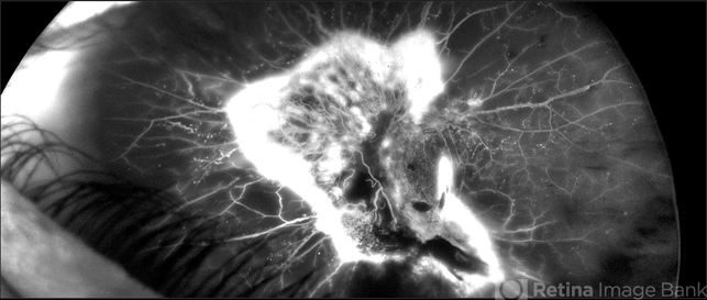
- 28-year-old white male with poorly controlled Type 1 DM, with a history of non-compliance with follow-ups, was referred with for DR with CME OS, and 3 weeks decrease vision OS. Va cc was 20/15 OD and HM OS. IOP 18/14. No NVI OU. Posteriorly, the right eye had macular exudates, no NVD, and a large area of NVE along the IT arcade. The left eye large NV plaque around disc, wrapping macula, with total RD with a posterior funnel appearance. The FA in the left eye showed severe peripheral and macular ischemia with diffuse leakage from a fibrovascualr plaque.


---thumb.jpg/image-square;max$79,0.ImageHandler "PDR with TRD and FVP")
---thumb.jpg/image-square;max$79,0.ImageHandler "Progressive BDR/ PDR")
---thumb.jpg/image-square;max$79,0.ImageHandler "Progressive BDR/ PDR")
---thumb.jpg/image-square;max$79,0.ImageHandler "Progressive BDR/ PDR")
---thumb.jpg/image-square;max$79,0.ImageHandler "Progressive BDR/ PDR")
---thumb.jpg/image-square;max$79,0.ImageHandler "Progressive BDR/ PDR")


