 Initializing download.
-
 By Aditya S Kelkar, MS, FRCS, FASRS,FRCOphth
NATIONAL INSTITUTE OF OPHTHALMOLOGY
Co-author(s): Dr. Jai Kelkar. Dr. Vikrant Narwadr - Uploaded on Nov 12, 2019.
- Last modified by Caroline Bozell on Nov 14, 2019.
- Rating
- Appears in
- Miscellaneous
- Condition/keywords
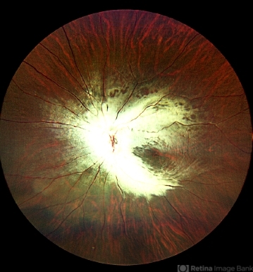
- myelinated nerve fibers
- Photographer
- Dr. Vikrant Narwade
- Imaging device
- Fundus camera
- Description
- Left eye fundus photograph of a 29-year-old male with extensive myelinated retinal nerve fibers appearing as grey white opaque lesions on the retina with feathery edges obscuring the retina.


---thumb.jpg/image-square;max$79,0.ImageHandler "Myelinated Nerve Fiber Layer")


---thumb.jpg/image-square;max$79,0.ImageHandler "Myelinated Nerve Fiber Layer")


