Fundus photograph of 43-year-old female with acute retinal necrosis
-
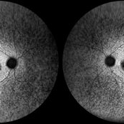
 Stargardt's Disease
Oct 4 2018 by Aditya S Kelkar, MS, FRCS, FASRS,FRCOphth
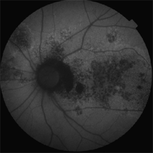
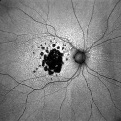
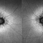
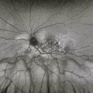
Auto-fluorescence image of a 19-year-old male showing flecks of both increased and decreased autofluorescence and reduced central macular autofluorescence surrounded by an increased signal.
Photographer: Dr. Aanchal Agarwal
Condition/keywords: Stargardt disease
-
 Chronic Multifocal Central Serous Chorio-Retinopathy
Jan 30 2019 by Aditya S Kelkar, MS, FRCS, FASRS,FRCOphth
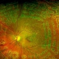
A 47-year-old male presented with left eye diminision of vision since 2 months. Left eye fundus autofluorescence image shows multiple hypofluorescent areas with numerous discrete small hyperfluroscent dots suggestive of inactive chronic multifocal central serous chorio-retinopathy.
Photographer: Dr. Abhishek Pandit
Condition/keywords: autofluorescence imaging, multifocal central serous chorioretinopathy (CSCR)
-
 Unilateral Myelinated Nerve Fiber
Nov 12 2019 by Aditya S Kelkar, MS, FRCS, FASRS,FRCOphth
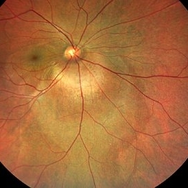
Left eye fundus photograph of a 29-year-old male with extensive myelinated retinal nerve fibers appearing as grey white opaque lesions on the retina with feathery edges obscuring the retina.
Photographer: Dr. Vikrant Narwade
Condition/keywords: myelinated nerve fibers
-
 Multifocal Choroiditis
Jul 19 2020 by Aditya S Kelkar, MS, FRCS, FASRS,FRCOphth
Fundus photograph of 29-year-old male with both eyes inactive multifocal choroiditis.
Photographer: Dr. Sayali Tidke
Imaging device: CLARUS 500
Condition/keywords: multifocal choroiditis
-
 Disciform Scar
Aug 18 2020 by Aditya S Kelkar, MS, FRCS, FASRS,FRCOphth
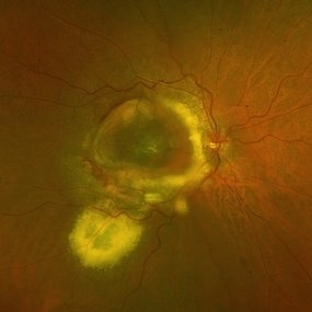
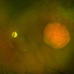
Left eye fundus photograph of 75-year-old male, showing large disciform scar post subretinal bleeding secondary to idiopathic polypoidal choroidal vasculopathy
Photographer: Dr.Mounika Bolisetty
Imaging device: CLARUS 500
Condition/keywords: disciform scar, idiopathic polypoidal choroidal vasculopathy
-
 Subhyaloid Hemorrhage
Aug 28 2020 by Aditya S Kelkar, MS, FRCS, FASRS,FRCOphth
Right eye fundus photograph of 56-year-old diabetic male , showing subhyaloid hemorrhage
Condition/keywords: subhyaloid hemorrhage
-
 Choroidal Disc Melanoma
Dec 24 2020 by Aditya S Kelkar, MS, FRCS, FASRS,FRCOphth
Melanoma or Melanocytoma- A mystery
Condition/keywords: melanocytoma
-
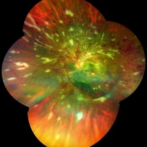
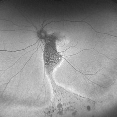
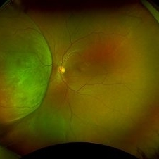
 Acute Retinal Necrosis
May 31 2021 by Aditya S Kelkar, MS, FRCS, FASRS,FRCOphth
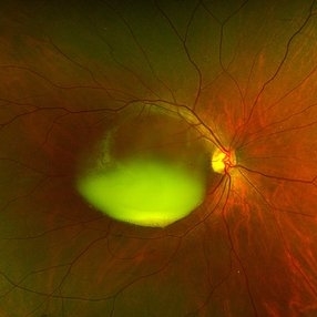
Fundus photograph of 43-year-old female with left eye acute retinal necrosis.
Imaging device: Clarus 500
Condition/keywords: acute retinal necrosis
-
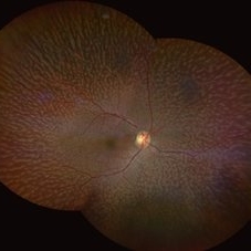
 Ocular Albinism
Aug 14 2021 by Aditya S Kelkar, MS, FRCS, FASRS,FRCOphth
Fundus photograph of the left eye of a 26-year-old young man with ocular albinism.
Photographer: Devesh Kumar Dahariya, National Institute of Ophthalmology, Pune, India.
Imaging device: Zeiss Clarus 500
Condition/keywords: ocular albinism
-
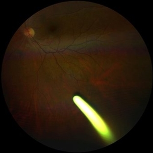
 Crystals in the Eye
Sep 3 2021 by Aditya S Kelkar, MS, FRCS, FASRS,FRCOphth
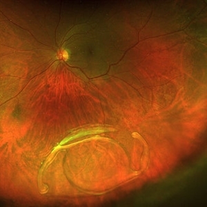
Left eye fundus photo of a 28 year-old , with air-filled vitreous cavity entering through the scleral wound site, after removal of impacted IOFB.
Imaging device: Clarus 500
Condition/keywords: intraocular foreign body, trauma
-
 Traumatic Retinal Tear
Dec 5 2021 by Aditya S Kelkar, MS, FRCS, FASRS,FRCOphth
Color fundus photograph of a 34-year old man's left eye, 2 hours after a tennis ball injury, showing commotio retinae with Berlin's edema and cherry red spot in the fovea along with linear retinal tears in the temporal equatorial zone.
Photographer: Dr Sukanya Mondal. National Institute of Ophthalmology, Pune. India.
Imaging device: Zeiss Clarus 500
Condition/keywords: Berlin's edema, cherry red spot, commotio retinae, retinal tear
-
 Paravenous-Pigmented-Retinochoroidal-Atrophy
Dec 17 2021 by Aditya S Kelkar, MS, FRCS, FASRS,FRCOphth
Right-eye Fundus Photo of a 30-year-old male.
Imaging device: Clarus 500
Condition/keywords: pigmented paravenous chorioretinal atrophy (PPCRA), retinochoroidopathy
-
 Traumatic Retinal Tear
Jan 20 2022 by Aditya S Kelkar, MS, FRCS, FASRS,FRCOphth
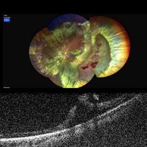
Color fundus photograph of a 34-year old man's left eye, 2 hours after a tennis ball injury, showing commotio retinae with Berlin's edema and cherry red spot in the fovea along with linear retinal tears in the temporal equatorial zone. Adjoining OCT slice taken through the break shows full thickness retinal tear without any underlying choroidal rupture.
Photographer: Dr Sukanya Mondal, National Institute of Ophthalmology, Pune. India
Imaging device: Zeiss Clarus 500
Condition/keywords: Berlin's edema, cherry red spot, commotio retinae, retinal tear
-
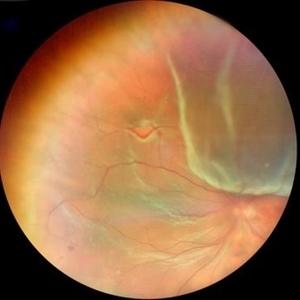
 Coats' Disease
Mar 30 2022 by Aditya S Kelkar, MS, FRCS, FASRS,FRCOphth
Colour Fundus photograph of a 3-year-old boy presenting with complaints of slowly progressive divergent squint of his left eye which on funduscopy revealed presence of Coats' Disease in the same eye.
Photographer: Dr. Pranali Surawase. National Institute of Ophthalmology, Pune, Maharashtra, India
Imaging device: Zeiss Clarus 500
Condition/keywords: angioma, cholesterol crystals, Coats' disease
-
 Stargardt disease
May 16 2022 by Aditya S Kelkar, MS, FRCS, FASRS,FRCOphth
15- year old, presented with blurring of vision since 10 years.
Photographer: Dr Mounika Bolisetty
Imaging device: CLARUS 500
Condition/keywords: macular dystrophy
-
 Fundus Albipunctatus
Aug 25 2022 by Aditya S Kelkar, MS, FRCS, FASRS,FRCOphth
65 year old female, presented for cataract evaluation. Fundus examination showed whitish-yellow flecks in the retina.
Imaging device: Clarus 500
Condition/keywords: fundus albipunctatus, fundus photograph
-
 Optic Disc Coloboma
Aug 27 2022 by Aditya S Kelkar, MS, FRCS, FASRS,FRCOphth
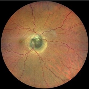
Color fundus photograph of a 51-year-old man showing optic disc coloboma of the left eye.
Photographer: Dr. Sukanya Mondal. National Institute of Ophthalmology, Pune, India.
Imaging device: Zeiss Clarus 500
Condition/keywords: coloboma of optic disc, color fundus photograph
-
 Retinal Detachment
Oct 3 2022 by Aditya S Kelkar, MS, FRCS, FASRS,FRCOphth
Middle aged female presented with blurring of vision in the right eye since 3 days. Fundus examination showed presence of retinal detachment with horse shoe tear
Imaging device: Clarus- 500
Condition/keywords: retinal tear
-
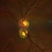
 Pseudo Foster Kennedy Syndrome
Oct 13 2022 by Aditya S Kelkar, MS, FRCS, FASRS,FRCOphth
Colour fundus photograph of a 44-year-old man showing bilateral small discs with optic atrophy on the right eye and disc edema on the left eye resulting from consecutive NAAION in both eyes.
Photographer: Dr Sukanya Mondal, National Institute of Ophthalmology, Pune. India
Imaging device: Zeiss Clarus 500
Condition/keywords: ischemic optic neuropathy, optic atrophy, optic disc edema
-
 Familial Exudative Vitreoretinopathy
Nov 25 2022 by Aditya S Kelkar, MS, FRCS, FASRS,FRCOphth
Colour fundus photograph of the right eye of a 56-year-old lady showing lasered FEVR with epiretinal membrane and vitreous band.
Photographer: Dr. Pranali Surawase. National Institute of Ophthalmology, Pune, Maharashtra, India
Imaging device: Zeiss Clarus 500
Condition/keywords: ERM, familial exudative vitreoretinopathy (FEVR), laser photocoagulation
-
 Idiopathic Vasculitis
Feb 4 2023 by Aditya S Kelkar, MS, FRCS, FASRS,FRCOphth
Color fundus photograph of the left eye showing idiopathic retinal vasculitis.
Photographer: Dr. Pranali Surawase. National Institute of Ophthalmology, Pune, India.
Imaging device: Zeiss Clarus 500
Condition/keywords: retinal vasculitis, vasculitis
-
 Intravitreal Ozurdex Implant
Feb 6 2023 by Aditya S Kelkar, MS, FRCS, FASRS,FRCOphth
Color fundus photograph of left eye showing intravitreal ozurdex implant
Photographer: Dr. Tanya Sagar. National institute of Ophthalmology, Pune, India.
Imaging device: Zeiss Clarus 500
Condition/keywords: dexamethasone implant, intravitreal implant, Ozurdex implant
-
Iatrogenic Macular Hole and Subretinal Migration of PFCL
Feb 7 2023 by Aditya S Kelkar, MS, FRCS, FASRS,FRCOphth
The video demonstrates a surgical scenario where the fovea gives away by the force imparted by the jet of an injecting PFCL (Perfluorocarbon heavy Liquid) and the PFCL migrates subfoveally. Intraoperative OCT confirms the presence of a macular hole. The situation is managed by ILM peeling and mobilizing subfoveal PFCL peripherally by injecting another bubble of PFCL over the posterior pole. A peripheral drainage retinotomy is then created to aspirate the subretinal PFCL followed by fluid-air exchange, PFCL-air exchange, and endolaser around the retinotomy. Post-operative OCT at 3 weeks’ follow-up shows a sealed macular hole.
Condition/keywords: Iatrogenic macular hole, Intraoperative complications, Subretinal PFCL
-
 RETINAL DETACHMENT WITH CHOROIDAL COLOBOMA
Feb 8 2023 by Aditya S Kelkar, MS, FRCS, FASRS,FRCOphth
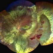
Left eye color fundus photograph of a 24 year old male showing retinal detachment with choroidal coloboma.
Photographer: Dr. Apoorva Jadhav , National Institute of Ophthalmology, Pune, India.
Imaging device: Zeiss Clarus 500
Condition/keywords: choroidal coloboma
-
 Choroidal-Nevus
Feb 25 2023 by Aditya S Kelkar, MS, FRCS, FASRS,FRCOphth
Color fundus photograph of the left eye of a 55 year old male showing large choroidal nevus.
Photographer: Dr. Apoorva Jadhav, National Institute of Ophthalmology, Pune. India.
Imaging device: Zeiss Clarus 500
Condition/keywords: Choroidal nevus
-
 Rhegmatogenous Retinal Detachment
Feb 26 2023 by Aditya S Kelkar, MS, FRCS, FASRS,FRCOphth
Color fundus photograph of left eye showing rhegmatogenous retinal detachment.
Photographer: Dr. Sahil Wagh, National Institute of Ophthalmology, Pune, India.
Imaging device: Zeiss Clarus 500
Condition/keywords: Retinal Detachment, rhegmatogenous retinal detachment
-
 Choroidal Melanoma with Exudative Retinal Detachment
Mar 2 2023 by Aditya S Kelkar, MS, FRCS, FASRS,FRCOphth
Color fundus photograph of the left eye of a 45 year old male showing choroidal melanoma with exudative retinal detachment.
Photographer: Dr. Pranali Surawase, National Institute of Ophthalmology, Pune, India.
Imaging device: Zeiss Clarus 500
Condition/keywords: choroidal mass, exudative retinal detachment, Retinal detachment
-
 LEBER'S MILIARY ANEURYSM
Mar 3 2023 by Aditya S Kelkar, MS, FRCS, FASRS,FRCOphth
Color fundus photograph of the left eye of a 53 year old female with leber's miliary aneurysm.
Photographer: Dr. Tanya Sagar, National Institute of Ophthalmology, Pune, India.
Imaging device: Zeiss Clarus 500
Condition/keywords: aneurysm, lipid exudation
-
 Inferior retinal detachment with lattice and holes
May 31 2023 by Aditya S Kelkar, MS, FRCS, FASRS,FRCOphth
Importance of dilated retina check up before Lasik surgery can't be better demonstrated...patient totally asymptomatic came for Lasik opinion and has inferior retinal detachment with lattice and holes, sparing the macula
Photographer: Dr. Sahil Wagh , National Institute of Opthalmology, Pune , India
Imaging device: Zeiss Clarus 500
Condition/keywords: inferior retinal detachment
-
 Rescuing IOL CTR Bag Complex
Jun 14 2023 by Aditya S Kelkar, MS, FRCS, FASRS,FRCOphth
INTRAOPERATIVE SNAPSHOPT IN ZEISS ARTEVO 800 OF DROPPED IOL CTR BAG COMPLEX IN A 71 YEAR OLD MALE PATIENT
Photographer: SUBHASREE DUTTA, NATIONAL INSTITUTE OF OPHTHALMOLOGY, PUNE
Imaging device: ZEISS ARTEVO 800
Condition/keywords: dropped capsular IOL bag complex
-
 RESCUING IOL CTR BAG COMPLEX
Jun 14 2023 by Aditya S Kelkar, MS, FRCS, FASRS,FRCOphth
INTRAOPERATIVE SNAPSHOPT IN ZEISS ARTEVO 800 OF DROPPED IOL CTR BAG COMPLEX IN A 71 YEAR OLD MALE PATIENT
Photographer: SUBHASREE DUTTA, NATIONAL INSTITUTE OF OPHTHALMOLOGY, PUNE
Imaging device: ZEISS ARTEVO 800
Condition/keywords: dropped capsular IOL bag complex
-
 Solitary large Congenital Hypertrophy of Retinal Pigment Epithelium (CHRPE)
Jul 1 2023 by Aditya S Kelkar, MS, FRCS, FASRS,FRCOphth
Right eye fundus photograph of a 42 year old asymptomatic male demonstrating a superotemporal solitary large Congenital Hypertrophy of Retinal Pigment Epithelium (CHRPE) lesion.
Photographer: Optom Komal Jangam
Imaging device: OPTOS DAYTONA
Condition/keywords: CHRPE
-
 Pseudo-doubling of optic nerve with Coloboma
Jul 10 2023 by Aditya S Kelkar, MS, FRCS, FASRS,FRCOphth
Right eye fundus photograph of a 41 year old asymptomatic female demonstrating Pseudo-doubling of optic nerve with Coloboma.
Photographer: Miss Komal Jangam,B.Sc Optometry, National Institute of Ophthalmology, Pune, India.
Imaging device: OPTOS DAYTONA
Condition/keywords: coloboma, Pseudoduplication of optic disc
-
 Optic Disc Drusen Autofluorescence
Jul 16 2023 by Aditya S Kelkar, MS, FRCS, FASRS,FRCOphth
Fundus autofluorescence imaging of a 22-year-old male with optic disc drusen seen as hyperautofluorescent spot.
Photographer: Dr. Harsh Jain
Condition/keywords: Autofluorescence imaging of Optic Disc Drusen
-
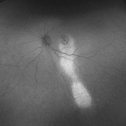 Comet Tail Sign In Chronic Central Serous Chorioretinopathy
Jul 23 2023 by Aditya S Kelkar, MS, FRCS, FASRS,FRCOphth
Fundus Autofluorescence imaging of an 57-year-old woman with Chronic Central Serous Chorioretinopathy demonstrating a Comet Tail Sign.
Photographer: Dr. Ajinkya Rawale. National institute of Ophthalmology, Pune, India.
Imaging device: OPTOS DAYTONA
Condition/keywords: chronic central serous chorioretinopathy (CSCR)
-
 Optic Disc Pit OCT
Aug 1 2023 by Aditya S Kelkar, MS, FRCS, FASRS,FRCOphth
Optical Coherence Tomography of an 21 year old male with a Optic Disc Pit.
Photographer: Dr. Ajinkya Rawale. National institute of Ophthalmology, Pune, India.
Imaging device: Zeiss Plex
Condition/keywords: congenital optic nerve pit
-
 Chorioretinal Coloboma
Aug 7 2023 by Aditya S Kelkar, MS, FRCS, FASRS,FRCOphth
Fundus photograph of an 68-year-old woman with a chorioretinal coloboma observed.
Photographer: Optom Komal Jangam, National Institute of Ophthalmology, Pune, India.
Imaging device: OPTOS DAYTONA
Condition/keywords: chorioretinal coloboma
-
 AGE RELATED MACULAR DEGENERATION AUTOFLUORESCENCE
Aug 13 2023 by Aditya S Kelkar, MS, FRCS, FASRS,FRCOphth
Autofluorescence fundus photography of an 78-year-old woman diagnosed with age-related macular degeneration.
Photographer: Dr. Harsh Jain, National Institute of Ophthalmology
Imaging device: Clarus 500
Condition/keywords: age-related macular degeneration (AMD)
-
 Endolaser in Status-Post Vitrectomy
Aug 28 2023 by Aditya S Kelkar, MS, FRCS, FASRS,FRCOphth
Endolaser in Status-Post Vitrectomy.
Photographer: Optom Komal Jangam, National Institute of Ophthalmology, Pune, India.
Imaging device: OPTOS DAYTONA
Condition/keywords: endolaser, pars plana vitrectomy (PPV), vitrectomy
-
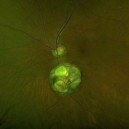
 Circumscribed Choroidal Hemangioma with Serous Macular Retinal Detachment
Oct 2 2023 by Aditya S Kelkar, MS, FRCS, FASRS,FRCOphth
Fundus photograph of a 43-year-old male with a circumscribed choroidal hemangioma with serous macular retinal detachment associated with diminision of vision.
Photographer: Dr. Harsh Jain, National Institute of Ophthalmology
Imaging device: Clarus 500
Condition/keywords: choroidal hemangioma
-
 DISCIFORM SCAR AND RETINAL PIGMENT EPITHELIUM (RPE) DETACHMENT IN A CASE OF IDIOPATHIC POLYPOIDAL CHOROIDAL VASCULOPATHY (IPCV)
Oct 21 2023 by Aditya S Kelkar, MS, FRCS, FASRS,FRCOphth
Right eye fundus photograph of a 83 year old female demonstrating Disciform Scar And Retinal Pigment Epithelium (RPE) Detachment In A Case Of Idiopathic Polypoidal Choroidal Vasculopathy (IPCV).
Photographer: DR APURVA MUKADAM
Imaging device: OPTOS DAYTONA
Condition/keywords: disciform scar
-
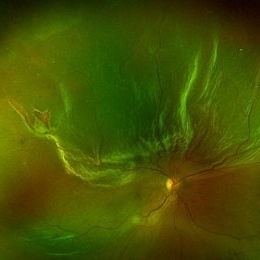
 Advanced Retinitis Pigmentosa
Mar 29 2024 by Aditya S Kelkar, MS, FRCS, FASRS,FRCOphth
Fundus photograph of an 70-year-old man with advanced retinitis pigmentosa.
Photographer: Optom Ayesha Inamdar, National Institute of Ophthalmology, Pune
Imaging device: Daytona OPTOS
Condition/keywords: RETINITIS PIGMENTOSA, RP
-
 Central Serous Chorioretinopathy With Comet Tail Sign
Jul 13 2024 by Aditya S Kelkar, MS, FRCS, FASRS,FRCOphth
Fundus Autofluorescence imaging of an 59-year-old man with Chronic Central Serous Chorioretinopathy demonstrating a Comet Tail Sign.
Photographer: Dr. Rabia Naaz, National institute of Ophthalmology, Pune, India
Imaging device: OPTOS DAYTONA
Condition/keywords: central serous chorioretinopathy (CSCR)
-
 Retinal Detachment with Giant Retinal Tear
Aug 9 2024 by Aditya S Kelkar, MS, FRCS, FASRS,FRCOphth
Fundus photograph of an 12-year-old boy with a Retinal detachment with Giant retinal tear of acute onset.
Photographer: Sakshi Naik, National Institute of Ophthalmology
Imaging device: Optos Daytona
Condition/keywords: giant retinal tear, pediatric retina, Retina detachment
-
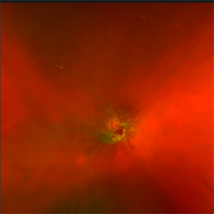 Ripples in Bloody Sea
Sep 14 2024 by Aditya S Kelkar, MS, FRCS, FASRS,FRCOphth
Fundus photograph of a 42-year-old man with a vitreous hemorrhage, with prominent neovascularization at disc.
Photographer: Neha Sharma
Imaging device: OPTOS DAYTONA
Condition/keywords: NEOVASCULARIZATION AT DISC, Vitreous hemorrhage
-
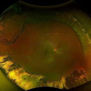 Status Post Scleral Buckle
Dec 9 2024 by Aditya S Kelkar, MS, FRCS, FASRS,FRCOphth
Fundus photograph of an 46-year-old man with status post scleral buckle in the right eye done 6 years ago.
Photographer: Optometrist Chandrakanta Bhandare, National Institute of Ophthalmology, Pune, India
Imaging device: OPTOS DAYTONA
Condition/keywords: Retinal Detachment, scleral buckle
-
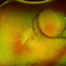
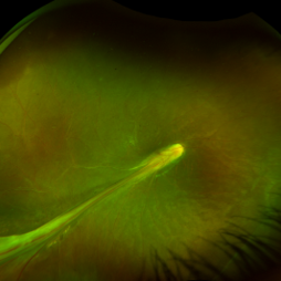 Falciform Retinal Detachment
Dec 24 2024 by Aditya S Kelkar, MS, FRCS, FASRS,FRCOphth
Fundus image of a 13 year-old female with falciform retinal detachment from the optic disc to the inferior-temporal QUADRANT
Photographer: NIKITA PABSETWAR, National institute of Ophthalmology
Imaging device: OPTOS DAYTONA
Condition/keywords: FALCIFORM RETINAL DETACHMENT, persistent fetal vasculature (PFV)
-
 Familial Exudative Vitreoretinopathy
Jan 17 2025 by Aditya S Kelkar, MS, FRCS, FASRS,FRCOphth
A fundus photograph of a 16-year-old boy reveals temporal peripheral retinal non-perfusion and incomplete vascularization.
Photographer: Optom Mansi Raut
Imaging device: Optos Daytona
Condition/keywords: familial exudative vitreoretinopathy (FEVR)
-
 Rod Cone Dystrophy
Mar 1 2025 by Aditya S Kelkar, MS, FRCS, FASRS,FRCOphth
Fundus Autofluorescence photograph of a 72-year-old woman with a rod cone dystrophy.
Photographer: Optom Rutuja Shelke
Imaging device: OPTOS DAYTONA
Condition/keywords: dystrophy
-
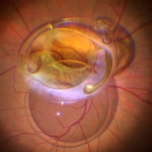
 Dislocated Intraocular Lens
Jun 4 2025 by Aditya S Kelkar, MS, FRCS, FASRS,FRCOphth
Fundus photograph of a 79-year-old man with a posteriorly dislocated intraocular lens in the inferior quadrant.
Photographer: Optom Chandrakanta Bhandare, National Institute of Ophthalmology, Pune
Imaging device: Optos Daytona
Condition/keywords: dislocated intraocular lens (IOL)
-
 Amelanotic Choroidal Melanoma with Optic Atrophy
Jun 11 2025 by Aditya S Kelkar, MS, FRCS, FASRS,FRCOphth
Fundus photograph of a 64-year-old woman with optic atrophy and amelanotic choroidal melanoma temporal to the macula.
Photographer: Dr Harsh Jain, National Institute of Ophthalmology
Imaging device: Optos Daytona
Condition/keywords: amelanotic melanoma, optic atrophy
-
 Chronic Sub-Hyaloid Hemorrhage with Dehemoglobinized Blood
Jul 11 2025 by Aditya S Kelkar, MS, FRCS, FASRS,FRCOphth
Fundus photograph of an 38-year-old man with a long standing sub hyaloid hemorrhage with dehemoglobinized blood.
Photographer: Optom Salomi Sonawane, National Institute of Ophthalmology, Pune, India
Imaging device: Optos Daytona
Condition/keywords: chronic, dehemoglobinized hemorrhage, SUBHYALOID HEMORRHAGE
-
 Exudative Retinal Detachment
Aug 6 2025 by Aditya S Kelkar, MS, FRCS, FASRS,FRCOphth
Fundus auto-fluorescence of a 41 year old female depicting retinal pigment epitheliopathy and exudative retinal detachment in case of ocular metastasis secondary to breast carcinoma.
Photographer: Dr.Rabia Naaz, National Institute of ophthalmology, Pune
Imaging device: OPTOS DAYTONA
Condition/keywords: Exudative retinal detachment, Retinal pigment epitheliopathy
-
 Dystrophy of the Retinal Pigment Epithelium
Aug 21 2025 by Aditya S Kelkar, MS, FRCS, FASRS,FRCOphth
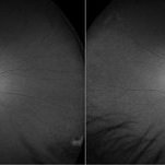
Both eyes autofluorescence imaging on Optos of a 56 year old female with complaints of gradual painless blurring of vision, aggravating on near work. Her BCVA for distance vision is 6/12 and 6/9 on snellens charting for Right and Left eye respectively. What could be the exact pathology or diagnosis? Kindly discuss and suggest.
Photographer: Dr. Muskan Mangal
Condition/keywords: autofluorescence imaging, Dystrophy of the Retinal Pigment Epithelium, macula lesion
-
 Adult Foveomacular Dystrophy
Aug 21 2025 by Aditya S Kelkar, MS, FRCS, FASRS,FRCOphth
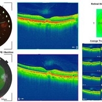
Left eye OCT of a 56 year old female with complaints of gradual painless blurring of vision, aggravating on near work.
Photographer: Dr. Muskan Mangal
Condition/keywords: adult foveomacular dystrophy, OCT
-
 Dystrophy of the Retinal Pigment Epithelium
Aug 21 2025 by Aditya S Kelkar, MS, FRCS, FASRS,FRCOphth
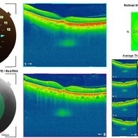
Right eye OCT of a 56 year old female with complaints of gradual painless blurring of vision, aggravating on near work.
Photographer: Dr. Muskan Mangal
Condition/keywords: Dystrophy of the Retinal Pigment Epithelium
-
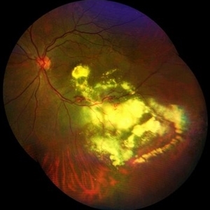
 Healed AZOOR- Multiple White Dot Syndrome
Aug 29 2025 by Aditya S Kelkar, MS, FRCS, FASRS,FRCOphth
Fundus photograph of a 58 year old woman with multiple, well-defined, punched-out chorioretinal scars scattered throughout the posterior pole and mid-periphery. The macular area shows a large, confluent, yellowish-white scar involving the fovea.
Photographer: Dr. Muskan Mangal
Imaging device: Optos Daytona
Condition/keywords: multifocal choroiditis, multiple evanescent white dot syndrome (MEWDS), punctate inner choroidopathy (PIC)
-
 Amelanotic Melanoma
Sep 19 2025 by Aditya S Kelkar, MS, FRCS, FASRS,FRCOphth
Widefield fundus photograph of a 37 year old showing a large, dome-shaped, intraocular mass involving the temporal retina. The lesion appears elevated and lacks surface pigmentation. Overlying retinal vessels are displaced and draped across the tumor surface, with surrounding retinal elevation noted. The appearance is suggestive of amelanotic variant of choroidal melanoma.
Photographer: Dr. Muskan Mangal
Imaging device: Optos Daytona
Condition/keywords: choroidal melanoma, intraocular tumor
-
 Rhegmatogenous Retinal Detachment
Sep 20 2025 by Aditya S Kelkar, MS, FRCS, FASRS,FRCOphth
A 56-year-old patient diagnosed with rhegmatogenous retinal detachment presents with two distinct retinal breaks observed on fundus imaging.
Photographer: Dr. muskan
Imaging device: optos daytona
Condition/keywords: rhegmatogenous retinal detachment, superior retina
-
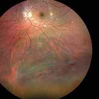
 Intricate Dance of Hemorrhage: BRVO with SHH with VH
Oct 11 2025 by Aditya S Kelkar, MS, FRCS, FASRS,FRCOphth
Retinal image of a 31-year-old male diagnosed with Branch Retinal Vein Occlusion (BRVO) alongside a subhyaloid hemorrhage and vitreous hemorrhage. The BRVO is evident by the disrupted blood flow in the retinal veins, leading to fluid leakage and hemorrhages. The blood leakage here shows the intricate pattern of hemorrhage revealing the hidden secrets of ocular health.
Photographer: Rhishita
Imaging device: optos daytona
Condition/keywords: branch retinal vein occlusion (BRVO), Hemorraghe, Retinal Vein Occlusion, Sub hyaloid haemorrhage, Vitreous hemorrhage
-
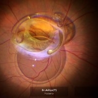
 Posterior Dislocated Intraocular Lens
Oct 23 2025 by Aditya S Kelkar, MS, FRCS, FASRS,FRCOphth
Fundus photograph of a 53-year-old man with a posteriorly dislocated intraocular lens near the posterior pole.
Photographer: Dr Tejal Rao, National Institute of Ophthalmology, Pune, India
Imaging device: Optos Daytona
Condition/keywords: dislocated intraocular lens (IOL), IOL drop