 Initializing download.
-
 By Lancaster Course in Ophthalmology
1980 - Uploaded on Mar 4, 2019.
- Last modified by Caroline Bozell on Mar 14, 2019.
- Rating
- Appears in
- Unit 08 Pathology of the Vitreous
- Condition/keywords
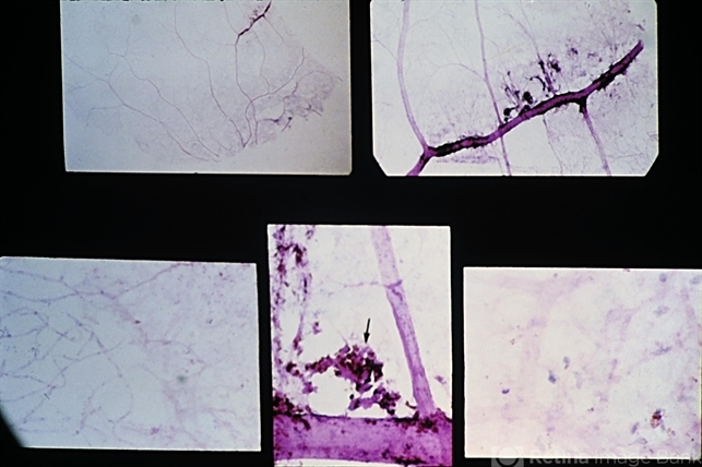
- lattice degeneration, nodular proliferations, sclerotic vessels, retinal pigment epithelium, endothelial, intramural pericytes
- Description
- Radial perivascular lattice retinal degeneration. Lower left view shows abrupt transition of the normal capillary bed (left) and a sclerotic occluded capillary bed in the area of the lattice (right). Lower right view, under higher magnification, shows the loss of intramural pericytes and endothelial cells in the capillary bed of the area of lattice degeneration. Clumps of hyperplastic and migrated retinal pigment epithelium with nodular proliferations of basement membrane (arrow) are adherent to the sclerotic vessels. (E.P. No. 31493)


