-
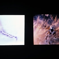
 Slide 8-1
Mar 4 2019 by Lancaster Course in Ophthalmology
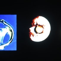
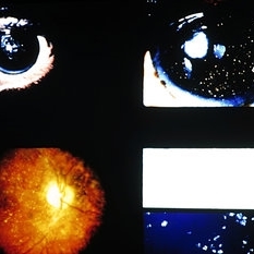
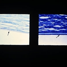
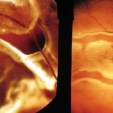
Left: Gross appearance of a funnel-shaped detachment of the vitreous. It remains attached at its base and at the optic nerve head. Center and right: Appearance of ring-shaped vitreous condensation, formerly attached at the optic nerve head. Such condensations can account for the visual symptom of floaters. (E.P. No. 21811)
Condition/keywords: detachment, operculum, vitreous
-
 Slide 8-2
Mar 4 2019 by Lancaster Course in Ophthalmology
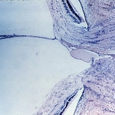
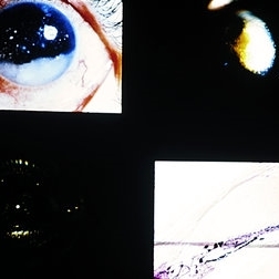
Hyaloid artery extending into the vitreous cavity from the optic nerve head. (E.P. No. 16127)
Condition/keywords: hyaloid artery, optic nerve head, vitreous cavity
-
 Slide 8-3
Mar 4 2019 by Lancaster Course in Ophthalmology
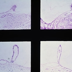
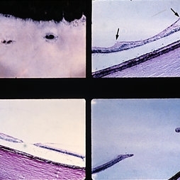
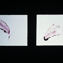
Examples of Bergmeister's papilla. Note the strands of vitreous, composed of glial cells, attached to the papilla. The presence of a small vessel within the Bergmeister's papilla (lower right) suggests that its origin is related to the hyaloid artery. (E. P. Nos. 31888,38589, 29360, and 32239)
Condition/keywords: Bergmeister's Papillae, glial cells, vitreous
-
 Slide 8-4
Mar 4 2019 by Lancaster Course in Ophthalmology
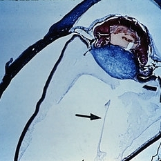
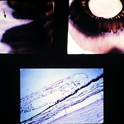
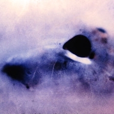
Persistence and hyperplasia of the primary vitreous (PHPV). The dense fibrovascular tissue molds the posterior surface of the lens and has drawn the ciliary processes and peripheral retina toward the center of the mass. A section of the hyaloid artery is present in the vitreous cavity (arrow). (A.F.l.P. No. 744398)
Condition/keywords: ciliary, fibrovascular tissue, hyaloid artery, persistent hyperplastic primary vitreous (PHPV)
-
 Slide 8-5
Mar 4 2019 by Lancaster Course in Ophthalmology
Left: Retinal pits along a major retinal vessel. (E.P. No. 31030) Right: Posterior detachment of the vitreous is shown, with several retinal pits and a small horseshoe-shaped retinal tear. (E.P. No. 30448)
Condition/keywords: posterior detachment, retinal pits, retinal tear, vitreous
-
 Slide 8-6
Mar 4 2019 by Lancaster Course in Ophthalmology
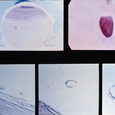
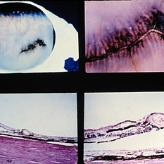
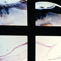
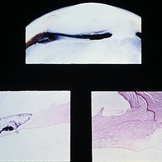
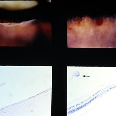
Gross and microscopic appearance of a retinal hole. Vitreous tractim has resulted in the hole and detachment of the operculum. Upper right shows the plug of retinal tissue (arrow) attached to the posterior aspect of the detached vitreous. Lower left shows the rounded posterior margin of the retinal hole and the small area of associated retinal detachment. Lower middle and right views are of sections through the detached retinal operculum. (E.P. No. 35238)
Condition/keywords: operculum, retinal hole, retinal tissue
-
 Slide 8-7
Mar 4 2019 by Lancaster Course in Ophthalmology
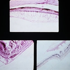
Meridional retinal fold. The peripheral retina is tented-up in a meridional direction (arrow) and is continuous with a dentate process. Lower view illustrates the retina extending over the pars plana. (E.P. No. 30600)
Condition/keywords: retinal fold
-
 Slide 8-8
Mar 4 2019 by Lancaster Course in Ophthalmology
Localized vitreous traction inducing cystic retinal degeneration and retinal detachment. (E.P. No. 19650)
Condition/keywords: cystic retinal degeneration, vitreous traction
-
 Slide 8-9
Mar 4 2019 by Lancaster Course in Ophthalmology
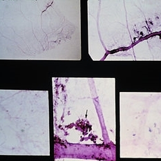
Lattice degeneration of retina with retinal holes. Microscopic appearance at three levels (upper, right, and lower) shows discontinuity of the internal limiting membrane, an overlying pocket of fluid vitreous, atrophy of inner retinal layers, hole formation, and vitreoretinal adhesion (arrows) at margin of lesion. (E.P. No. 30544)
Condition/keywords: atrophy, retinal degeneration, retinal hole, vitreoretinal adhesion
-
 Slide 8-10
Mar 4 2019 by Lancaster Course in Ophthalmology
Gross appearance of an area of lattice degeneration with vitreous traction and hole formation at the posterior margin. (E.P. No. 30755)
Condition/keywords: lattice degeneration
-
 Slide 8-11
Mar 4 2019 by Lancaster Course in Ophthalmology
Radial perivascular lattice degeneration. The lesion is located at or posterior to the equator and is associated with a major vessel. As with a typical lattice there is discontinuity of the internal limiting membrane, a loss of inner retinal layers, an overlying vitreous degeneration, vitreoretinal adhesion at the margin, sclerosis of vessels, and retinal pigment epithelial hypertrophy, hyperplasia, and migration into the retina. (E.P. No. 31493)
Condition/keywords: hyperplasia, lattice degeneration, retinal pigment epithelium, vitreoretinal adhesion, vitreoretinal degeneration
-
 Slide 8-12
Mar 4 2019 by Lancaster Course in Ophthalmology
Radial perivascular lattice retinal degeneration. Lower left view shows abrupt transition of the normal capillary bed (left) and a sclerotic occluded capillary bed in the area of the lattice (right). Lower right view, under higher magnification, shows the loss of intramural pericytes and endothelial cells in the capillary bed of the area of lattice degeneration. Clumps of hyperplastic and migrated retinal pigment epithelium with nodular proliferations of basement membrane (arrow) are adherent to the sclerotic vessels. (E.P. No. 31493)
Condition/keywords: endothelial, intramural pericytes, lattice degeneration, nodular proliferations, retinal pigment epithelium, sclerotic vessels
-
 Slide 8-13
Mar 4 2019 by Lancaster Course in Ophthalmology
Asteroid hyalosis. These opacities may be polychromatic. Lower right view shows the histologic appearance and the birefringence of these opacities.
Condition/keywords: asteroid hyalosis, birefringence, opacities
-
 Slide 8-14
Mar 4 2019 by Lancaster Course in Ophthalmology
Cholesterosis bulbi (hemophthalmos). When in the anterior chamber, these crystals have a whitish or yellowish-gold appearance. Upper right and lower views are of the same case (E.P. No. 31377) and show cholesterol in the anterior and posterior chambers.
Condition/keywords: cholesterolosis bulbi, hemophthalmos
-
 Slide 8-15
Mar 4 2019 by Lancaster Course in Ophthalmology
Retinal detachment due to vitreous incarceration in the cataract wound and traction on the retina inferiorly. The tented-up retina has been pulled over the pars plana. (E.P. No. 32421)
Condition/keywords: cataract, inferiorly, pars plana, vitreous
-
 Slide 8-16
Mar 4 2019 by Lancaster Course in Ophthalmology
Updrawn pupil following cataract extraction. Vitreous is incarcerated in the cataract scar above. The inferior iris leaf is adherent to fibrous tissue that has proliferated along the tract of the anterior vitreous. Contraction of this fibrosed anterior vitreous has pulled the attached inferior iris leaf toward the wound and has resulted in an updrawn pupil. (E.P. No. 31006)
Condition/keywords: cataract, cataract extraction, fibrous tissue, iris leaf, updrawn pupil, vitreous
-
 Slide 8-17
Mar 4 2019 by Lancaster Course in Ophthalmology
Right: Aphakic bullous keratopathy with vitreous touch. The endothelium is markedly flattened with extreme irregular spacing of nuclei. One flattened endothelial-cell nucleus is present (arrow) (E.P. No. 39964) Left: Aphakic bullous keratopathy with vitreous touch, partial endothelial atrophy, and retrocorneal fibrous tissue proliferation (arrow). (E.P. No. 39234)
Condition/keywords: atrophy, keratopathy, retrocorneal fibrous tissue proliferation, vitreous touch
-
 Slide 8-18
Mar 4 2019 by Lancaster Course in Ophthalmology
lridovitreous synechia at pupillary margin (arrow, left) and at iridectomy site (arrow, right) in an eye following cataract extraction. (E.P. No. 32655)
Condition/keywords: cataract extraction, synechiae
-
 Slide 8-19
Mar 4 2019 by Lancaster Course in Ophthalmology
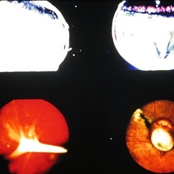
Traumatic retinal dialysis in a 15-year-old male who died 2 weeks following a motorcycle accident. Upper left shows dialysis of the retina at the ora serrata. Upper right shows hemorrhage In the retina of the fellow eye. Sections through dialysis (lower views) show the detached vitreous base with a small tag of adherent retinal tissue (arrow). (E.P. No. 32955)
Condition/keywords: hemorrhage, ora serrata, retinal dialysis
-
 Slide 8-20
Mar 4 2019 by Lancaster Course in Ophthalmology
Examples of fibrous tissue proliferation in the vitreous at sites of penetration of globe. Fibrous encasement of a retained intraocular foreign body is illustrated in the lower right view.
Condition/keywords: fibrous tissue proliferation
-
 Slide 8-21
Mar 4 2019 by Lancaster Course in Ophthalmology
Retinal tears at or near major retinal vessels. The vessel bridging the retinal hole (left) may be the source of vitreous hemorrhage. (Courtesy of E. D. Norton, M.D.)
Condition/keywords: retinal hole, retinal tear, vitreous hemorrhage
-
 Slide 8-22
Mar 4 2019 by Lancaster Course in Ophthalmology
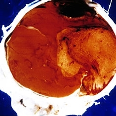
Extensive vitreous and intraocular hemorrhage from a necrotic malignant melanoma. The eye was enucleated with the diagnosis of absolute glaucoma and possible old central retinal vein occlusion. (E.P. No. 31366)
Condition/keywords: intraocular hemorrhage, melanoma, vitreous hemorrhage
-
 Slide 8-24
Mar 4 2019 by Lancaster Course in Ophthalmology
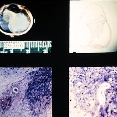
Diffuse nematode endophthalmitis with a fibroinflammatory mass in the vitreous (upper views) with eosinophils and the nematode larva, and total retinal detachment. (E.P. No. 29566)
Condition/keywords: endophthalmitis, fibroinflammatory mass
-
 Slide 8-25
Mar 4 2019 by Lancaster Course in Ophthalmology
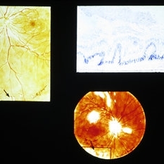
Composite showing various lesions due to sarcoidosis. The drawings show seeding of sarcoid nodules in the vitreous (arrows). Histologic section through such a lesion shows granulomatous inflammatory aggregates extending into the vitreous from the retina (upper right).
Condition/keywords: lesions, sarcoid nodules, sarcoidosis
-
 Slide 8-26
Mar 4 2019 by Lancaster Course in Ophthalmology
Primary amyloidosis with amyloid deposits in vitreous adjacent to the retina (left). (Courtesy of R. G. Michels, M. D.)
Condition/keywords: amyloidosis
-
 Slide 8-27
Mar 4 2019 by Lancaster Course in Ophthalmology
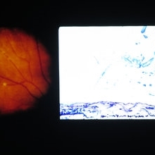
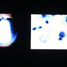
Ocular reticulum-cell sarcoma. Appearance of vitreous cells and debris (left) in a 61-year-old man with a 2-year history of recalcitrant uveitis. Pars plana vitrectomy specimen was prepared, using a millipore filter and staining by a modified Papanicolaou technique. Tumor cells with nuclear cytoplasmic disproportion and finger like nuclear extensions are characteristic of reticulum-cell sarcoma (right). (E.P. No. 38606)
Condition/keywords: pars plana vitrectomy (PPV), reticulum cell sarcoma, uveitis, vitreous cells
-
 Slide 8-28
Mar 4 2019 by Lancaster Course in Ophthalmology
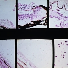
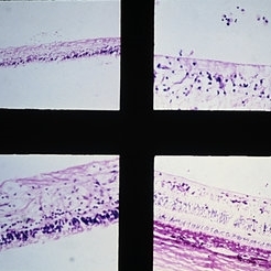
Reticulum-cell sarcoma. Right eye of a 74-year-old female with a history of bilateral uveitis for 1 year. The right eye became blind and painful and was enucleated 6 months later. A mild nongranulomatous inflammatory infiltrate was present in the iris and ciliary body (upper views). Inflammatory cells and tumor cells were found in the vitreous (lower views). (E.P. No. 36437)
Condition/keywords: nongranulomatous inflammatory infiltrate, reticulum cell sarcoma, uveitis
-
 Slide 8-29
Mar 4 2019 by Lancaster Course in Ophthalmology
Whipple's disease with inflammatory cells in the vitreous and marked histiocytic infiltration in the retina (arrow). These histiocytes contain PAS-positive material (lowe right). (A.F.I.P. No. 1548417)
Condition/keywords: histiocytic infiltration, inflammatory cells, PAS positive material, Whipple's disease
A project from the American Society of Retina Specialists