25 year old woman with 7 days history of central vision loss and photopsias
-
 Multifocal Choroiditis
Aug 16 2018 by FELIPE PEREIRA
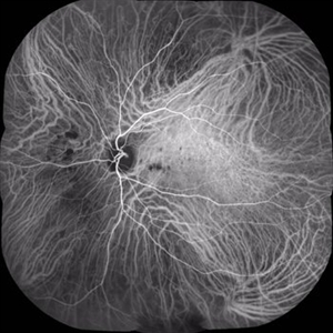
Mid-phase indocyanine green angiography of a 25-year-old woman with sudden central vision loss and photopsias for 7 days. The hypofluorescent lesions in the macula and nasal to the disc correspond to the yellow-white deep lesions in the fundus examination. No leakage is observed at any stage of the exam
Photographer: Claudio Zett Lobos
Imaging device: HEIDELBERG SPECTRALIS HRA
Condition/keywords: indocyanine green (ICG) angiography, multifocal choroiditis, white dot syndrome
-
 Multifocal Choroiditis
Dec 22 2018 by FELIPE PEREIRA
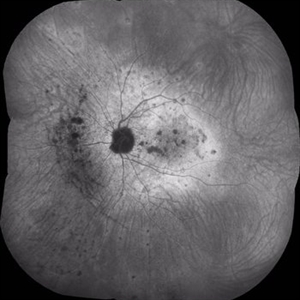
Late phase ICG exam of a 25-year-old woman with 7 days history of central vision loss and photopsias. The hypofluorescent dots corresponds to the classically yellow-white lesion in the retinography. In the late fases the hypofluorescent lesions acquire a hiperfluorescent ring of staining. This image demonstrate more lesion than is possible to see in the clinical exam.
Photographer: Claudio Zett Lobos
Imaging device: HEIDELBERG SPECTRALIS HRA
Condition/keywords: indocyanine green (ICG) angiography, multifocal choroiditis, white dot syndrome