 Initializing download.
-
 By Dhaivat Shah
Sankara Nethralaya
Co-author(s): Dr Eesh Nigam, Dr Purna Nangia - Uploaded on Sep 21, 2018.
- Last modified by Caroline Bozell on Sep 21, 2018.
- Rating
- Appears in
- Imaging marvels
- Condition/keywords
- polypoidal choroidal vasculopathy (PCV)
- Photographer
- Miss Moupiya Das
- Imaging device
- Scanning laser ophthalmoscope
- Description
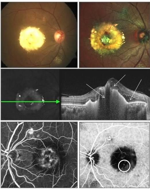
- A 40-year-old female presented with sudden onset decreased vision in right eye. BCVA: CF 1 mt. Fundus showed massive subretinal exudation with haemorrhage. EDI OCT showed notched PEDs with shallow SRF and exudation with back-shadowing. FFA shows leak with window defects. ICG shows hotspot in late phase. Polypoidal choroidal vasculopathy (PCV) is a retinal disorder characterized by the presence of aneurysmal polypoidal lesions in the choroidal vasculature, resulting in damage to the overlying retina and loss of retinal pigment epithelium. The aneurysmal dilatations, also known as polyps, may be found subfoveal, juxtafoveal, extrafoveal, peripapillary or even peripheral regions. The polypoidal lesions are best detected on indocyanine green angiography as hotspots in late phase. The presence of choroidal polyps can lead to recurrent episodes of exudative retinal detachment, serous or hemorrhagic pigment epithelial detachment, subretinal hemorrhage and exudation. Treatment is available in form of laser/PDT along with Anti VEGF injection.


---thumb.jpg/image-square;max$79,0.ImageHandler "Polypoidal Choroidal Vasculopathy: Case 1 - Image 1 of 7")
---thumb.jpg/image-square;max$79,0.ImageHandler "Polypoidal Choroidal Vasculopathy: Case 1 - Image 3 of 7")

---thumb.jpg/image-square;max$79,0.ImageHandler "Polypoidal Choroidal Vasculopathy: Case 1 - Image 2 of 7")


---thumb.jpg/image-square;max$79,0.ImageHandler "Polypoidal Choroidal Vasculopathy - Case 1")