 Initializing download.
-
 By Hosam Attia, MD
- Uploaded on Jun 1, 2018.
- Last modified by Caroline Bozell on Jun 5, 2018.
- Rating
- Appears in
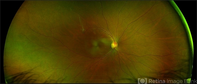
- Tractional Vs Combined TractionalRhegmatogenous Retinal Detachment with active Neovascularization
- Condition/keywords
- neovascularization elsewhere (NVE), tractional retinal detachment, combined retinal detachment
- Imaging device
-
Scanning laser ophthalmoscope
Optos California - Description
- 47-year-old African American, with history of diabetes mellitus of unknown duration and control, was referred for initial evaluation for conjunctival laceration in his left eye, following accidental finger nail injury, 6 days prior to presentation. - On exam, his vision was 20/50 OD and Bare HM/ LP OS. - Fundus color photos OD: No significant pathology, aside from attenuated vasculature OS: Chronic, Mac-Off, almost closed funnel tractional vs combined tractional/rhegmatogenous retinal detachment with large neovascularization (NVE) superiorly, detached ghost vessels, mild fresh vitreous hemorrhage, sub-retinal bands and inferior white vitreous debris from old hemorrhage (Not shown) - FA OD: No significant pathology aside from possible mild capillary non-perfusion in the extreme periphery, attenuated vasculature and possible tiny microaneurysms, nasally. OS: Extensive, wide spread capillary non- perfusion (correlate w/ detached Ghost vessels on color photos), and leakage from the NVE. - B/L Carotid Duplex was recommended due to the striking asymmetry in pathology with unknown medical history, diabetes duration and control, etc (even in absence of any signs suggestive of possible ocular ischemic syndrome OD)


---thumb.jpg/image-square;max$79,0.ImageHandler "Complications of ARN, TRD")

