-
 Retinal Detachment
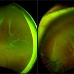
May 15 2018 by Morgan Benton
Ultra-wide field pseudocolor image of a 54-year-old male with a retinal detachment affecting his left eye after trauma. Patient was only able to see hand motion.
Photographer: Morgan Benton
Imaging device: Optos
Condition/keywords: color photo, left eye, Optos, ultra-wide field imaging
-
 Proliferative Diabetic Retinopathy
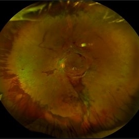
May 15 2018 by Morgan Benton
Ultra-wide field pseudocolor image of a 42-year-old female with proliferative diabetic retinopathy resulting in severe hemorrhaging. Vision was cc20/80+1 when the image was taken.
Photographer: Morgan Benton
Imaging device: Optos
Condition/keywords: color fundus photograph, hemorrhage, left eye, montage, neovascularization (NV), Optos, proliferative diabetic retinopathy (PDR), ultra-wide field imaging
-
 Proliferative Diabetic Retinopathy
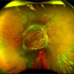
May 15 2018 by Morgan Benton
Ultra-wide field pseudocolor image of a 42-year-old female with proliferative diabetic retinopathy resulting in a tractional retinal detachment. Vision was cc20/50-2+1 when the image was taken.
Photographer: Morgan Benton
Imaging device: Optos
Condition/keywords: color fundus photograph, neovascularization (NV), Optos, proliferative diabetic retinopathy (PDR), tractional retinal detachment, ultra-wide field imaging
-
 Dislocated IOL
May 15 2018 by Morgan Benton
Ultra-wide field pseudocolor image of a 68-year-old male with a dislocated IOL after cataract surgery in the left eye. Patient was only able to count fingers at one foot and could pinhole to 20/60.
Photographer: Morgan Benton
Imaging device: Optos
Condition/keywords: color fundus photograph, dislocated intraocular lens (IOL), left eye, Optos, ultra-wide field imaging
A project from the American Society of Retina Specialists