File number: 28154
Comments
-
 Vishal Agrawal, MD, FRCS,FACS,FASRS (June 11 2018)
Thank you very much .
-
 Suber S. Huang, MD, MBA, FASRS (June 7 2018)
Full marks. Thank you for sharing this spectacular image!
Sign in to comment.
 Initializing download.
-
 By Vishal Agrawal, MD, FRCS,FACS,FASRS
Agrawal Hospital , Jaipur
Co-author(s): Dr Kamlesh Khilnani - Uploaded on Apr 30, 2018.
- Last modified by Caroline Bozell on Jul 6, 2018.
- Image of the week
-
Jul 8, 2018
View all images of the week - Rating
- Appears in
- 30-Apr-2018
- Condition/keywords
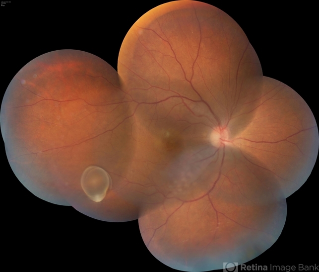
- cysticercosis, full thickness macular hole
- Photographer
- Vishal Agrawal MD,FRCS
- Imaging device
-
Fundus camera
Zeiss 524 - Description
- Fundus montage picture of a 40-year-old man presenting with decreased vision in the right eye for the past 2 months. Live intravitreal cysticercosis can be seen lying on the retina. Zooming the image reveals the full thickness macular hole. The scolex invaginates with the light of the camera causing double image of the cyst because of movement .


