-
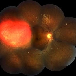
 Giant Retinal Tear-Retinal Detachment
May 25 2017 by Somnath Chakraborty, MD
Fundus photograph of a 45-year-old female with pathological myopia and retinal detachment secondary to giant retinal tear.
Photographer: Saptarshi Mehta, Retina Institute of Bengal
Condition/keywords: giant retinal tear, retinal tear
-
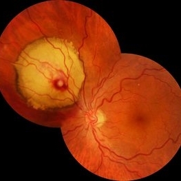
 Supero-Temporal Bullous Retinal Detachment With Macular Splitting
Sep 15 2017 by Somnath Chakraborty, MD
Montage fundus photo of a 56-year-old female with a bullous rhegmatogenous retinal detachment in the supero-temporal quadrant, secondary to a large horse shoe tear at 10 o' clock hour. She also has a large, pigmented lattice extending from 4 to 6 o' o' clock hours.
Photographer: Saptarshi Mehta, Retina Institute of Bengal
Condition/keywords: macular splitting, pigmented lattice lesion, retinal tear
-
 Multiple Retinal Lesions Secondary to Blunt Trauma
Jun 19 2018 by Somnath Chakraborty, MD
A montage of the right eye of a 15-year-old boy, who was struck by a football. The image shows multiple choroidal ruptures in the macular area, with sub-retinal blood and multiple, large retinal tears temporally. There is also an area of juxtapapillary, pigmentary changes.
Photographer: Saptarshi Mehta, Retina Institute of Bengal
Condition/keywords: blunt trauma, choroidal rupture, giant retinal tear, subretinal hemorrhage
-
 Focal Chroidal Hemangioma
Sep 18 2018 by Somnath Chakraborty, MD
Right eye fundus photo montage of a 17-year-old boy showing a focal choridal hemangioma temporally.
Photographer: Saptarshi Mehta, Retina Institute of Bengal
Condition/keywords: choroidal hemangioma
-
 Retinal Arterial Macroaneurysm
Sep 18 2018 by Somnath Chakraborty, MD
Left eye fundus photo of a 72-year-old hypertensive, female with a hemorrhagic retinal arterial macroaneurysm with sub-retinal blood.
Photographer: Pulok Chandra Roy, Retina Institute of Bengal
Condition/keywords: retinal arterial macroaneurysm
-
 Intraocular Foreign Body
Feb 7 2019 by Somnath Chakraborty, MD
Left eye fundus photo montage of a 45-year-old male showing a large iron foreign body, impacted inferior to the infero-temporal branch vessels with a large patch of surrounding chorio-retinal atrophy, secondary to resolving Commotio retinae
Photographer: Saptarshi Mehta
Condition/keywords: commotio retinae, intraocular foreign body, trauma
-
 Retinal CRAO With Emboli
Jun 27 2019 by Somnath Chakraborty, MD
Left eye fundus photo montage of a 43-year-old male with central retinal artery occlusion with bright yellow multiple retinal (cholesterol) emboli both at the disc and also along multiple retinal arteries.
Photographer: Pulak Roy
Condition/keywords: arterial embolus, central retinal artery occlusion (CRAO), cholesterol embolus
-
 Diabetic Macular TRD
Jan 10 2020 by Somnath Chakraborty, MD
Fundus Montage image of the left eye of a 48-year-old type 2 diabetic with post PRP macular extensive tractional retinal detachment involving macula.
Photographer: Pulak Roy
Condition/keywords: diabetic retinopathy, proliferative diabetic retinopathy (PDR), tractional retinal detachment, vitrectomy, vitreomacular surgery
-
 Occlusive Retinal Vasculitis
Dec 17 2020 by Somnath Chakraborty, MD
Fundus photo montage of left eye of a 45-year-old male showing a large neovascular frond secondary to peripheral occlusive vasculitis.
Photographer: Pulak Roy
Condition/keywords: neovascularization elsewhere (NVE), peripheral retinal vasculitis, retinal vasculitis
-
 RD Montage
Jul 3 2021 by Somnath Chakraborty, MD
Fundus photo montage of the left eye of a 56-year-old male showing subtotal retinal detachment with macular involvement and a large circumlinear tear extending from 1 o' clock to 3 o' clock hours.
Photographer: Pulak Roy
Condition/keywords: acute retinal detachment, retinal detachment of the macula, retinal tear, retinal tear with detachment
-
 High risk Proliferative Diabetic Retinopathy treated with Pan Retinal Photocoagulation
Nov 5 2022 by Somnath Chakraborty, MD
A Fundus Photo Montage of 43 year old Asian Male with Type 2 Diabetes Mellitus since 7 years who presented with sudden onset diminition of vision in his Left eye. BCVA OS was 20/200. He was diagnosed to have Pre retinal bleed due to Proliferative Diabetic Retinopathy and was treated with Pan Retinal Photocoagulation. This image shows a large neo-cascular frond at the disc and superior to it with Pre-retinal bleed and Fresh laser marks along
Photographer: Pulak Roy
Condition/keywords: diabetic blindness, diabetic retinopathy vitrectomy study (DRVS), fresh laser burns, laser photocoagulation, preretinal hemorrhage, proliferative diabetic retinopathy (PDR)
A project from the American Society of Retina Specialists