File number: 26764
Comments
-
 Yuntao Hu, MD, PhD (March 29 2018)
It is better there is a picture of B scan for this eye
Sign in to comment.
 Initializing download.
-
 By Guruprasad S. Ayachit, MBBS,MS
M.M.JOSHI EYE INSTITUTE
Co-author(s): Shrinivas M Joshi, M M Joshi Eye Institute, Hubli; Apoorva Ayachit, M M Joshi Eye Institute, Hubli - Uploaded on Oct 5, 2016.
- Last modified by Caroline Bozell on Nov 23, 2016.
- Image of the week
-
Nov 27, 2016
View all images of the week - Rating
- Appears in
- Miscellaneous
- Condition/keywords
- combined hamartoma
- Photographer
- Shravan Masurkar, M M Joshi Eye Institute, Hubli
- Imaging device
-
Fundus camera
Topcon TRC50DX - Description
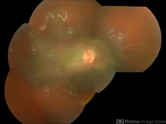
- Fundus photograph of a 9-year-old boy with an ill-defined lesion extending from nasal to the disc going on to include the papillomacular bundle; 14X10 mm in greatest dimensions. There is a thick epiretinal membrane causing distortion and straightening of temporal vascular arcade.
---thumb.jpg/image-square;max$79,0.ImageHandler "Combined Hamartoma")


