File number: 26497
Comments
-
 James B. Soque, CRA, OCT-C, COA, FOPS (August 29 2016)
Excellent montage Olivia! Your focus of the inferior arcade of this right eye is exceptional. Thank you for this superb submission.
Sign in to comment.
 Initializing download.
-
 By Olivia Rainey
Retina Specialists of Michigan
Co-author(s): Thomas Aaberg Jr., MD - Uploaded on May 22, 2016.
- Last modified by Olivia Rainey on May 5, 2020.
- Rating
- Appears in
- Miscellaneous
- Condition/keywords
- arteriovenous malformation, montage, color fundus photograph, color photo, peripheral ischemia
- Photographer
- Olivia Rainey
- Imaging device
-
Fundus camera
Topcon 50dx - Description
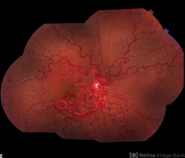
- Color fundus montage of an 13-year-old female with arteriovenous malformation (Wyburn Mason Racemose Angiomatosis) affecting her right eye. The retinal arteriovenous malformation appears to be stable. She presented with NLP in the eye, strabismus, and peripheral retinal ischemia. She is at risk for neovascular complications; however, she is currently being treated with Sirolimus. Since she is on this systemically, there is no need to perform intraocular anti-VEGF injections or PRP laser. She also presented with optic atrophy affecting her left eye, secondary to chiasmal involvement of arteriovenous malformation. She has had a potential progressive visual field loss involving the temporal aspect of her visual field from the left eye. There is sector optic atrophy. Presumably, this is due to a compressive effect of her arteriovenous malformation on the nasal nerve fiber layer (corresponding to the temporal visual field) crossing to the right occipital cortex at the chiasm.


