 Initializing download.
-
 By Mallika Goyal, MD
Apollo Health City
Co-author(s): Sridhar A, MD, Apollo Health City, Hyderabad, India - Uploaded on Feb 10, 2016.
- Last modified by Caroline Bozell on Feb 11, 2016.
- Rating
- Appears in
- Bilateral Myopic Foveoschisis
- Condition/keywords
- myopic foveoschisis
- Photographer
- Mallika Goyal, MD, Apollo Health City, Hyderabad, India
- Imaging device
- Fundus camera
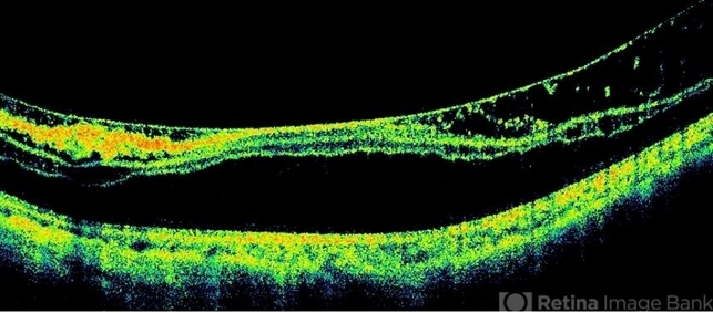
- Description
- Left eye OCT of a 22-year-old lady with bilateral myopic foveoschisis who presented with complaint of left eye vision drop 4 months prior to presentation. BCVA was 20/400. This OCT scan is taken inferior to foveal centre. The scan through foveal centre reveals an outer lamellar macular hole accounting for vision loss.
---thumb.jpg/image-square;max$79,0.ImageHandler "Myopic Traction Maculopathy")
---thumb.jpg/image-square;max$79,0.ImageHandler "Myopic Traction Maculopathy")


