-
 Bilateral Myopic Foveoschisis
Feb 10 2016 by Mallika Goyal, MD
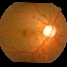
Right eye fundus of a 22-year-old lady with bilateral myopic foveoschisis. BCVA is 20/25, and she is visually asymptomatic in this eye.
Photographer: Mallika Goyal, MD, Apollo Health City, Hyderabad, India
Condition/keywords: myopic foveoschisis
-
 Bilateral Myopic Foveoschisis
Feb 10 2016 by Mallika Goyal, MD
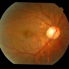
Right eye fundus of a 22-year-old lady with bilateral myopic foveoschisis. BCVA is 20/25, and she is visually asymptomatic in this eye.
Photographer: Mallika Goyal, MD, Apollo Health City, Hyderabad, India
Condition/keywords: myopic foveoschisis
-
 Bilateral Myopic Foveoschisis
Feb 10 2016 by Mallika Goyal, MD
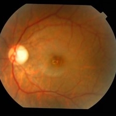
Right eye fundus of a 22-year-old lady with bilateral myopic foveoschisis. BCVA is 20/25, and she is visually asymptomatic in this eye.
Photographer: Mallika Goyal, MD, Apollo Health City, Hyderabad, India
Condition/keywords: myopic foveoschisis
-
 Bilateral Myopic Foveoschisis
Feb 10 2016 by Mallika Goyal, MD
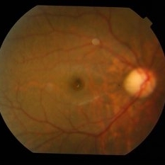
Left eye fundus of a 22-year-old lady with bilateral myopic foveoschisis who presented with complaint of left eye vision drop 4 months prior to presentation. BCVA was 20/400. OCT scan through foveal centre revealed an outer lamellar macular hole accounting for vision loss.
Photographer: Mallika Goyal, MD, Apollo Health City, Hyderabad, India
Condition/keywords: myopic foveoschisis
-
 Bilateral Myopic Foveoschisis
Feb 10 2016 by Mallika Goyal, MD
Right eye OCT of a 22-year-old lady with bilateral myopic foveoschisis. BCVA is 20/25, and she is visually asymptomatic in this eye.
Photographer: Mallika Goyal, MD, Apollo Health City, Hyderabad, India
Condition/keywords: myopic foveoschisis
-
 Bilateral Myopic Foveoschisis
Feb 10 2016 by Mallika Goyal, MD
Left eye OCT of a 22-year-old lady with myopic foveoschisis & an outer lamellar macular hole who presented with complaint of left eye vision drop 4 months prior to presentation. BCVA was 20/400. The lamellar macular hole accounts for the vision loss.
Photographer: Mallika Goyal, MD, Apollo Health City, Hyderabad, India
Condition/keywords: myopic foveoschisis
-
 Bilateral Myopic Foveoschisis
Feb 10 2016 by Mallika Goyal, MD
Left eye OCT of a 22-year-old lady with bilateral myopic foveoschisis who presented with complaint of left eye vision drop 4 months prior to presentation. BCVA was 20/400. This OCT scan is taken superior to foveal centre. The scan through foveal centre reveals an outer lamellar macular hole accounting for vision loss.
Photographer: Mallika Goyal, MD, Apollo Health City, Hyderabad, India
Condition/keywords: myopic foveoschisis
-
 Bilateral Myopic Foveoschisis
Feb 10 2016 by Mallika Goyal, MD
Left eye OCT of a 22-year-old lady with bilateral myopic foveoschisis who presented with complaint of left eye vision drop 4 months prior to presentation. BCVA was 20/400. This OCT scan is taken inferior to foveal centre. The scan through foveal centre reveals an outer lamellar macular hole accounting for vision loss.
Photographer: Mallika Goyal, MD, Apollo Health City, Hyderabad, India
Condition/keywords: myopic foveoschisis
A project from the American Society of Retina Specialists