-
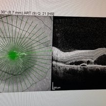 OCT
OCT
Apr 30 2015 by Mariam A Al-Feky, MD
A case of CSR phtographed on the Heidelberg fundus camera with multicolor image, infrared, blue reflectance and green reflectence predye injection, postdye injection and during dye injection and last image for the OCT. Fundus examination after dye injection showed a green spot nasal that was not detected predye injection. Multicolor image was retaken and that green spot is well evident in the multicolor image, the infrared relectance, blue and green reflectance. That green spot is corresponding to the leaky point in FFA and to a PED in OCT.
Photographer: Mariam AL-Feky
Condition/keywords: central serous retinopathy (CSR), leakage
-
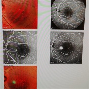 FFA
FFA
Apr 30 2015 by Mariam A Al-Feky, MD
A case of CSR phtographed on the Heidelberg fundus camera with multicolor image, infrared, blue reflectance and green reflectence predye injection, postdye injection and during dye injection and last image for the OCT. Fundus examination after dye injection showed a green spot nasal that was not detected predye injection. Multicolor image was retaken and that green spot is well evident in the multicolor image, the infrared relectance, blue and green reflectance. That green spot is corresponding to the leaky point in FFA and to a PED in OCT.
Photographer: Mariam AL-Feky
Condition/keywords: central serous retinopathy (CSR), leakage
-
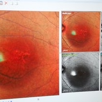 postinjection
Apr 30 2015 by Mariam A Al-Feky, MD
A case of CSR phtographed on the Heidelberg fundus camera with multicolor image, infrared, blue reflectance and green reflectence predye injection, postdye injection and during dye injection and last image for the OCT. Fundus examination after dye injection showed a green spot nasal that was not detected predye injection. Multicolor image was retaken and that green spot is well evident in the multicolor image, the infrared relectance, blue and green reflectance. That green spot is corresponding to the leaky point in FFA and to a PED in OCT.
Photographer: Mariam AL-Feky
Condition/keywords: central serous retinopathy (CSR), leakage
-
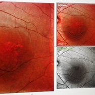 Central serous chorioretinopathy
Apr 30 2015 by Mariam A Al-Feky, MD
A case of CSR phtographed on the Heidelberg fundus camera with multicolor image, infrared, blue reflectance and green reflectence predye injection, postdye injection and during dye injection and last image for the OCT. Fundus examination after dye injection showed a green spot nasal that was not detected predye injection. Multicolor image was retaken and that green spot is well evident in the multicolor image, the infrared relectance, blue and green reflectance. That green spot is corresponding to the leaky point in FFA and to a PED in OCT.
Photographer: Mariam AL-Feky
Condition/keywords: central serous retinopathy (CSR), leakage
-
 Astrocytic Hamartoma
Apr 30 2015 by Mariam A Al-Feky, MD
A 15-year-old boy with history of seizures controlled on treatment. C/O: OD painless DV 10/7 ago (accidental discovery) O/E: BCVA OD: 6/60 ,, OS 6/6. AS: NAD OU. Pupil: RRR no RAPD OU. Fundus examination OD showed a retinitis like lesion with an overlying corkscrew vessel well evident on FFA with late leakage and CSR and OCT through the retinitis like lesion shows diffuse hypereflective thickeninig in the superficial NFL. Thorough history taking revealed that patient has seizures and MRI lesions suggestive of tuberous sclerosis. So this is exudative hamartoma secondary to tuberous sclerosis with marked resolution after single IVI of Lucentis. Retinitis like lesion with corkscrew vessels in FFA is typical together with the homogenous hypereflective thickening in the NFL.
Photographer: Mariam AL-Feky
Imaging device: Optical coherence tomography
Condition/keywords: astrocytic hamartoma
-
 Optic Nerve Head Drusen
Sep 10 2015 by Mariam A Al-Feky, MD
Right eye, 30-year-old obese female patient with BCVA 0.8 OU presenting with severe headache of 1 month duration. Ant seg: NAD OU, post. segment: disc edema OU, MRI brain is normal. ONH drusen is the diagnosis actually, with late staining in FFA (without early hyperfluorescent telangectatic disc capillaries as in caes of papillaedema), well delineated in the ONH map on the Heidelberg machine, and could be detected as a hypereflective material below the nerve fiber layer on the line scan. N.B. Burried ONH drusen don't autofluoresce.
Imaging device: Fundus camera
Condition/keywords: optic nerve head
-
 Optic Nerve Head Drusen
Sep 10 2015 by Mariam A Al-Feky, MD
Left eye 30-year-old obese female patient with BCVA 0.8 OU presenting with severe headache of 1 month duration Ant seg: NAD OU, post. segment: disc edema OU MRI brain is normal ONH drusen is the diagnosis actually, with late staining in FFA (without early hyperfluorescent telangectatic disc capillaries as in cases of papillaedema), well delineated in the ONH map on the Heidelberg machine, and could be detected as a hypereflective material below the nerve fiber layer on the line scan. N.B. Burried ONH drusen don't autofluoresce.
Imaging device: Fundus camera
Condition/keywords: drusen, optic nerve head
A project from the American Society of Retina Specialists