-
 Chronic Central Serous Chorioretinopathy (CSCR)
Nov 15 2014 by Rita Couceiro, MD, MS
53-year-old black male, with no relevant prior medical history, complained of bilateral blurry vision for the previous 16 years. On examination, visual acuity was 20/50 on the right eye (OD) and 20/100 on the left eye (OS). Anterior segment evaluation was unremarkable. Fundoscopy revealed pigmentary changes near the macular area in both eyes, with a mottling configuration, suggesting chronic CSCR. Fluorescein angiography showed an ink-blot pattern, with leakage superior to the fovea in OD and nasal to the fovea in OS.
Photographer: Telma Gala - Hospital de Santa Maria, Lisbon, Portugal
Condition/keywords: chronic central serous chorioretinopathy (CSCR)
-
 Chronical Submacular Hemorrhage in the Setting of Neovascular AMD
Mar 23 2015 by Rita Couceiro, MD, MS
An 80-year-old male, with a history of hypertension and high cholesterol, complained of acute and painless vision loss in his left eye (OS) in the previous 5 months. On observation best corrected visual acuity in OS was hand motion. A dense vitreous opacity in OS precluded fundus examination. Ocular ultrasound revealed vitreous hemorrhage and thickening of the macular area. The patient was submitted to pars plana vitrectomy, which disclosed a large submacular hemorrhage with chronical features and disciform scarring in the setting of neovascular AMD.
Imaging device: Intraoperative fundus photograph
Condition/keywords: neovascular age-related macular degeneration (AMD), submacular hemorrhage, wet age-related macular degeneration (wet AMD)
-
 Combined Hamartoma of the Retina and RPE
Apr 16 2015 by Rita Couceiro, MD, MS
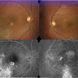
Fundus photograph, red-free picture (top images) and fluorescein angiography pictures (bottom images) of a 6-year-old girl with a combined hamartoma of the retina and RPE in the right eye.
Condition/keywords: hamartoma
-
 Coats Disease
Apr 16 2015 by Rita Couceiro, MD, MS
Fundus photograph and fluorescein angiography pictures of a 13-year-old girl with Coats Disease, showing abnormal telangiectatic vessels and intense exsudation in the inferior retinal periphery of the left eye.
Condition/keywords: Coats' disease, retinal telangiectasia
-
 Retinal Astrocytoma
Apr 16 2015 by Rita Couceiro, MD, MS
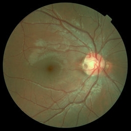
Fundus photograph of a 72-year-old man with a retinal astrocytoma of the left eye.
Condition/keywords: astrocytic hamartoma
-
 Retinal Pigment Changes After Blunt Ocular Trauma
Jun 27 2016 by Rita Couceiro, MD, MS
A 19-year-old man suffered blunt trauma of the left eye with a ball during soccer practice. At day 3 after trauma (upper pictures) the retinal area superior to the fovea looked pale and visual acuity was reduced to 20/32. This area revealed hypersignaling of retinal layers on OCT and the foveal area showed a localized disruption of retinal layers above the RPE. At day 30 (lower pictures) the retinal area of pallor showed pigmentary changes and OCT revealed atrophy of the external retinal layers. However the localized subfoveal retinal disruption was improved and only a slight disruption was seen on OCT at the ellipsoid level. Visual acuity of the left eye was restored to 20/20 although a scotoma remained.
Photographer: Rita Couceiro, Serviço de Oftalmologia do Hospital de Santa Maria, Lisboa, Portugal
Condition/keywords: blunt trauma, commotio retinae, pigment changes
-
 Optic Disc Pit
Nov 27 2016 by Rita Couceiro, MD, MS
15-year-old boy with an optic disc pit of the right eye (incidental finding during routine fundoscopy).
Photographer: Andreia Rocha
Condition/keywords: optic disc pit
A project from the American Society of Retina Specialists