 Initializing download.
-
 By Norman Byer
From Dr. Norman E. Byer’s “The Peripheral Retina in Profile” - Uploaded on Nov 9, 2012.
- Last modified by Suber S. Huang, MD, MBA, FASRS on Feb 11, 2013.
- Reviewed by Chayal Patel
- Rating
- Appears in
- Miscellaneous
- Condition/keywords
- retinal schisis detachment, outer layer hole, retinoschisis, intact inner layer, localized detachment of outer layer, secondary retinal detachment
- Description
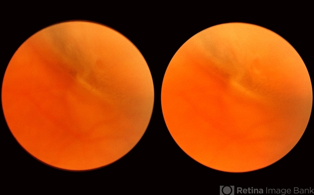
- This 57-year-old man has a combined retinal schisis detachment caused by an outer layer hole in the upper right. On the right half of this photograph, the outer layer is detached and represented by the prominent yellow line which is lying against the inner layer. On the left half the inner layer appears very thin and the schisis cavity lies just behind it as it was originally. This, therefore, represents a localized detachment of the outer layer and thus a true secondary retinal detachment. The reason these cases remain localized and nonprogressive is that the only fluid available to the subretinal space is that which is contained within the schisis cavity. Furthermore, this fluid tends to be quite viscous and is not readily passed through the retinal breaks. A clinical symptomatic progressive retinal detachment cannot occur unless the retinal schisis cavity is very large or a break occurs in the inner layer also.


")


---thumb.jpg/image-square;max$79,0.ImageHandler "Retinoschisis")


