 Initializing download.
-
 By Norman Byer
From Dr. Norman E. Byer’s “The Peripheral Retina in Profile” - Uploaded on Nov 9, 2012.
- Last modified by Suber S. Huang, MD, MBA, FASRS on Feb 11, 2013.
- Reviewed by Chayal Patel
- Rating
- Appears in
- Miscellaneous
- Condition/keywords
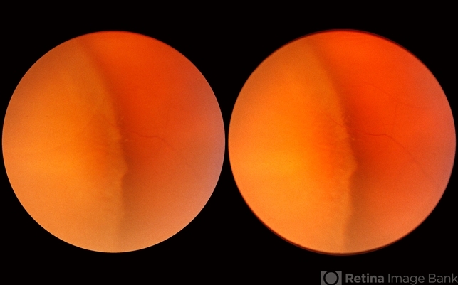
- retinoschisis, outer layer hole, yellow dots, intact inner layer, elevated outer layer, localized detachment of outer layer
- Description
- This is the same case as the previous two examples. The large outer layer hole of the previous photograph has led to a localized detachment of the outer layer. In this photograph, the camera is focused on the inner surface of the outer layer in order to show its irregular fur-like contour. Note also the tiny yellow dots of the inner layer superimposed on the dark indentation shadow. There is a fluid-filled space not only to the right but also to the left of the convex yellow line of the outer retinal layer indicating that the outer layer is elevated.


")


---thumb.jpg/image-square;max$79,0.ImageHandler "Retinoschisis")


