 Initializing download.
-
 By Norman Byer
From Dr. Norman E. Byer’s “The Peripheral Retina in Profile” - Uploaded on Nov 9, 2012.
- Last modified by Suber S. Huang, MD, MBA, FASRS on Feb 10, 2013.
- Reviewed by Chayal Patel
- Rating
- Appears in
- Miscellaneous
- Condition/keywords
- senile retinoschisis, yellow flecks, lower temporal quadrant
- Description
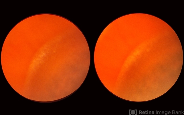
- This 48-year-old woman has senile retinoschisis involving the most common location, the lower temporal quadrant. The lesion shown here illustrates one of the two clinical features which are most often responsible for attracting the attention of the examiner to such lesions, namely the multitude of yellow flecks lying on the inner surface of the inner layer. The nature of these flecks is not known, but it seems clear that they do not originate in the schisis cavity for they do not represent remnants of ruptured Miller’s fibers. In this photograph you can easily detect the fluid space which separates the inner and outer retinal layers.


---thumb.jpg/image-square;max$79,0.ImageHandler "Stargardt's Dystrophy")


