 Initializing download.
-
 By Norman Byer
From Dr. Norman E. Byer’s “The Peripheral Retina in Profile” - Uploaded on Nov 9, 2012.
- Last modified by Suber S. Huang, MD, MBA, FASRS on Feb 10, 2013.
- Reviewed by Chayal Patel
- Rating
- Appears in
- Miscellaneous
- Condition/keywords
- lattice lesion, lattice degeneration, retinal hole, atrophic retinal hole, yellowish zone
- Description
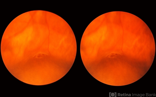
- In this 47-year-old woman, this lattice lesion with a small hole in the right end has led to a subclinical retinal detachment which extends to the margin of the subtle yellowish zone almost at the upper edge of the photograph. This patient did not desire surgery, and the area of detachment has changed only a small amount in the past eight years. The risk of a clinical retinal detachment developing from lattice degeneration is less than 1 percent. In those cases where it does though, about 3 quarters are caused by a tractional tear and about one quarter are caused by an atrophic hole as in this case.
---thumb.jpg/image-square;max$79,0.ImageHandler "Lattice Degeneration")


