 Initializing download.
-
 By Norman Byer
From Dr. Norman E. Byer’s “The Peripheral Retina in Profile” - Uploaded on Nov 9, 2012.
- Last modified by Suber S. Huang, MD, MBA, FASRS on Feb 10, 2013.
- Reviewed by Chayal Patel
- Rating
- Appears in
- Miscellaneous
- Condition/keywords
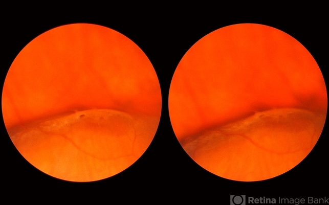
- glial tuft, lattice degeneration, round hole
- Description
- This 34 year-old man had a flat lattice lesion with no hole at this location for five years. Then he developed this round hole with a small subclinical retinal detachment which has not changed in appearance for four years. Note the tiny glial tuft just to the left of the hole and superimposed against the dark background.

---thumb.jpg/image-square;max$79,0.ImageHandler "Lattice Degeneration")


