 Initializing download.
-
 By Norman Byer
From Dr. Norman E. Byer’s “The Peripheral Retina in Profile” - Uploaded on Nov 9, 2012.
- Last modified by Suber S. Huang, MD, MBA, FASRS on Feb 10, 2013.
- Reviewed by Chayal Patel
- Rating
- Appears in
- Miscellaneous
- Condition/keywords
- posterior vitreous detachment, cystic retinal tuft, white retinal tuft, glial cells
- Description
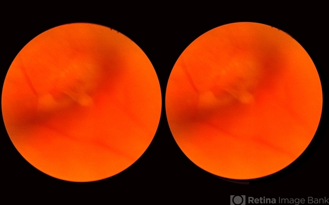
- This 68-year-old woman had a recent posterior vitreous detachment which produced this symptomatic horseshoe tear exactly at the site of this cystic retinal tuft. Note the characteristic discrete white nubbin at the apex, which is produced by a cap of glial cells with densely packed cytoplasm.


