 Initializing download.
-
 By Seif Allah Anwar
King Salman International University - Uploaded on Sep 16, 2025.
- Last modified by Joshua Friedman on Sep 17, 2025.
- Rating
- Appears in
- Miscellaneous
- Condition/keywords
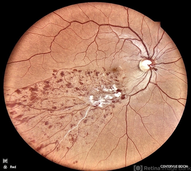
- Lower temporal branch retinal vein occlusion
- Photographer
- Dr Seif Anwar. FRCSEd
- Imaging device
- CENTERVUE EIDON
- Description
- Fundus photograph of a 46-year old hypertensive male patient showing sheathed lower temporal retinal vein with whitish cotton wool spots and hemorrhages ( dots, blots and flame shaped) along the area drained by the obstructed vein with vein.


")


