-
 Large Leaking Optic Disc Neovascularization
Sep 9 2025 by Seif Allah Anwar
Large leaking optic disc neovascularization.
Photographer: Dr Seif Anwar
Imaging device: Topcon
-
 Optic Disc Neovascularization
Sep 9 2025 by Seif Allah Anwar
A case of high risk proliferative diabetic retinopathy with large disc neovascularization.
Photographer: Dr Seif Anwar
Imaging device: Topcon
Condition/keywords: optic disc neovascularization
-
 Lower Temporal Branch Retinal Vein Occlusion
Sep 16 2025 by Seif Allah Anwar
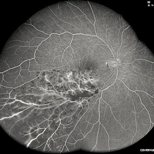
FFA of A 46-year old hypertensive male patient showing blocked fluorescence by retinal hemorrhages along the area drained by the obstructed vein with areas of retinal capillary drop-out.
Photographer: Dr Seif Anwar , FRCSEd
Imaging device: CENTERVUE EIDON
Condition/keywords: Lower temporal branch retinal vein occlusion
-
 Lower Temporal Branch Retinal Vein Occlusion
Sep 16 2025 by Seif Allah Anwar
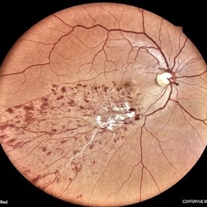
Fundus photograph of a 46-year old hypertensive male patient showing sheathed lower temporal retinal vein with whitish cotton wool spots and hemorrhages ( dots, blots and flame shaped) along the area drained by the obstructed vein with vein.
Photographer: Dr Seif Anwar. FRCSEd
Imaging device: CENTERVUE EIDON
Condition/keywords: Lower temporal branch retinal vein occlusion
-
 Chorioretinal Coloboma
Oct 6 2025 by Seif Allah Anwar
Fundus photograph of the patient left eye showing large, well-demarcated, excavated chorioretinal coloboma involving the inferior fundus, extending from the optic disc to the periphery. The lesion appears white due to bare sclera visibility, with absence of overlying choroid and retina. Retinal vessels course over the colobomatous area inferiorly.
Photographer: Dr. Seif Anwar, FRCSEd
Imaging device: Centervue Eidon
Condition/keywords: chorioretinal coloboma
-
 Dissociated Optic Nerve Fiber Layer (DONFL)
Oct 14 2025 by Seif Allah Anwar
54 year-old male patient, 7 months following PPV with ILM peeling for an idiopathic epiretinal membrane complaining of gradual painless diminution of vision starting 3 month following the operation , en face OCT at the inner retinal layer shows multiple concentric dark spots representing apoptosis of the RNFL and GCL.
Photographer: Dr.Seif Anwar, FRCSEd
Imaging device: Optovue, SOLIX OCT
Condition/keywords: En Face OCTA
-
 Racemose Hemangioma
Dec 16 2025 by Seif Allah Anwar
18 year-old female with dilated, tortuous arteriovenous communication without an intervening capillary bed. Vessels may appear coiled or spaghetti-like extending into the foveal region with no associated retinal hemorrhages, exudates, or edema On OCT : the anomalous vessels appear hyper reflective spanning the whole retinal thickness with ILM draping, No associated subretinal or intraretinal fluid.
Photographer: Seif Anwar , KING SALMAN INTERNATIONAL UNIVERSITY
Imaging device: TOPCON
Condition/keywords: racemose hemangioma
-
 Retinal Artery Macroaneurysms
Dec 29 2025 by Seif Allah Anwar
Multi Modal imaging ( color photo , OCT , OCT-A and en face OCTA ) of right lower temporal 3 adjacent leaking macro aneurysms in a 48 -year-old hypertensive female ( red arrows) with surrounding cystic oedema and exudations .
Photographer: Seif Anwar, FRCSEd
Imaging device: Fundus camera (TOPCON TRC 50X) & Optovue OCT (Solix)
Condition/keywords: Retinal artery Macroaneurysms
-
 Occlusive Retinal Vasculitis
Dec 30 2025 by Seif Allah Anwar
Ultra-wide field fundus photo of a 48 year old male patient with no history of hypertension or diabetes, presented with Marked perivascular sheathing, predominantly along the veins indicating active vasculitis (pattern resembling frosted branch angiitis) , with hemorrhages (dots , blots , flame shaped with dense pre macular hemorrhage) and hyperemic disc swelling.
Photographer: Seif Anwar, FRCSEd
Imaging device: Optos fundus Camera
Condition/keywords: occlusive retinal vasculitis
-
 Giant Tear Retinal Detachment
Jan 13 2026 by Seif Allah Anwar
Right eye funds photo of a 13 -year old boy following bluing ocular trauma showing giant retinal tear, the macula is off with the retinal flab reflected nasally.
Photographer: SeifAnwar, FRCSEd
Imaging device: Centervue Eidon
Condition/keywords: Giant tear retinal detachment
-
 Choroidal Rupture
Jan 16 2026 by Seif Allah Anwar
Wide crescent shaped choroidal rupture crossing the temporal foveal edge, in a 14 year child following blunt ocular trauma.
Photographer: Seif Anwar FRCSEd
Imaging device: Centervue Eidon
Condition/keywords: choroidal rupture
A project from the American Society of Retina Specialists