 Initializing download.
-
 By Felipe Murati
University of Arizona
Co-author(s): Juan B. Yepez, MD; Michele Petitto, MD; Igor Kozak, MD, PhD; J. Fernando Arevalo, MD, PhD - Uploaded on May 11, 2025.
- Last modified by Joshua Friedman on May 12, 2025.
- Rating
- Appears in
- Miscellaneous
- Condition/keywords
- Vogt-Koyanagi-Harada, pseudotumor, subretinal fibrosis, granulomatous uveitis, chronic inflammation, OCT
- Photographer
- Felipe A. Murati, MD, University of Arizona
- Imaging device
-
Fundus camera
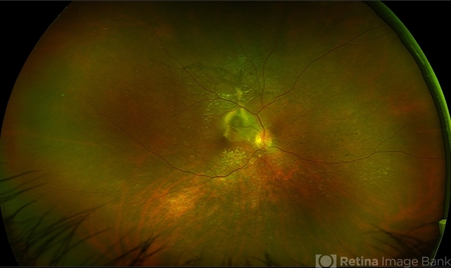
Optos California ultra-widefield retinal imaging system, single-capture, color fundus modality. - Description
- Ultra-widefield fundus image from a 36-year-old woman with chronic VKH syndrome showing a pseudotumor-like subretinal fibrotic lesion in the right eye. The lesion developed after multiple relapses and remained stable over a 1-year follow-up with immunosuppressive treatment including prednisone, mycophenolate mofetil, and adalimumab. No active choroiditis or exudative detachment was observed. Multimodal imaging was essential for disease monitoring.

")


---thumb.jpg/image-square;max$79,0.ImageHandler "Recurrent Pseudotumor with Scleritis")
---thumb.jpg/image-square;max$79,0.ImageHandler "Recurrent Pseudotumor with Scleritis")
---thumb.jpg/image-square;max$79,0.ImageHandler "Recurrent Pseudotumor with Scleritis")
---thumb.jpg/image-square;max$79,0.ImageHandler "Recurrent Pseudotumor")


