-
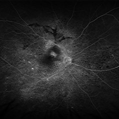 VKH Pseudotumor – Fluorescein Angiography
May 11 2025 by Felipe Murati
Fluorescein angiography image from a 36-year-old woman with chronic Vogt-Koyanagi-Harada (VKH) syndrome showing a pseudotumor-like lesion with late-phase staining and no active leakage. The image highlights subretinal fibrosis in the right eye, stable under long-term immunosuppressive therapy with mycophenolate mofetil and adalimumab. No signs of active choroiditis are present, confirming a quiescent phase.
Photographer: Felipe A. Murati, MD, University of Arizona
Imaging device: Optos California, fluorescein angiography modality
Condition/keywords: choroiditis, granulomatous uveitis, Optos FA, pseudotumor, subretinal fibrosis, VKH, Vogt-Koyanagi-Harada
-
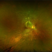 VKH Pseudotumor – Chronic Subretinal Fibrosis
May 11 2025 by Felipe Murati
Ultra-widefield fundus image from a 36-year-old woman with chronic VKH syndrome showing a pseudotumor-like subretinal fibrotic lesion in the right eye. The lesion developed after multiple relapses and remained stable over a 1-year follow-up with immunosuppressive treatment including prednisone, mycophenolate mofetil, and adalimumab. No active choroiditis or exudative detachment was observed. Multimodal imaging was essential for disease monitoring.
Photographer: Felipe A. Murati, MD, University of Arizona
Imaging device: Optos California ultra-widefield retinal imaging system, single-capture, color fundus modality.
Condition/keywords: adalimumab, chronic inflammation, granulomatous uveitis, OCT, Optos ultra-widefield imaging, pseudotumor, subretinal fibrosis, VKH, Vogt-Koyanagi-Harada
A project from the American Society of Retina Specialists