 Initializing download.
-
 By yuan duo
Co-author(s): Guoming Zhang Shenzhen Eye Hospital, Jinan University - Uploaded on Jan 16, 2025.
- Last modified by Joshua Friedman on Jan 17, 2025.
- Rating
- Appears in
- Miscellaneous
- Condition/keywords
- Retinal Ora Serrata
- Imaging device
- Scanning laser ophthalmoscope
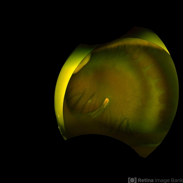
- Description
- A 5-year-old girl, born full-term with no history of systemic disease, presented with poor vision since early childhood and underwent fundus examination. Anterior segments of both eyes showed no significant abnormalities. Fundus examination revealed retinal folds extending from the optic disc to the temporal peripheral retina, with blood vessels coursing through the folds (A, B). Avascular zones were observed in the peripheral retina, and the ora serrata’s boundaries were clearly visible, displaying dentate processes and bays (C, D). Retinal pigmentation was evident. Genetic testing confirmed the final diagnosis of bilateral Familial Exudative Vitreoretinopathy (FEVR).


