-
 Familial Exudative Vitreoretinopathy
Jan 16 2025 by yuan duo
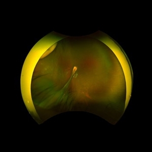
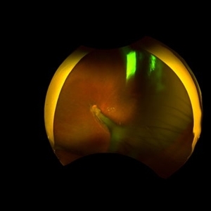
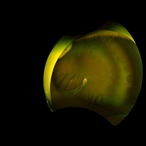
A 5-year-old girl, born full-term with no history of systemic disease, presented with poor vision since early childhood and underwent fundus examination. Anterior segments of both eyes showed no significant abnormalities. Fundus examination revealed retinal folds extending from the optic disc to the temporal peripheral retina, with blood vessels coursing through the folds (A, B). Avascular zones were observed in the peripheral retina, and the ora serrata’s boundaries were clearly visible, displaying dentate processes and bays (C, D). Retinal pigmentation was evident. Genetic testing confirmed the final diagnosis of bilateral Familial Exudative Vitreoretinopathy (FEVR).
Condition/keywords: Retinal Ora Serrata
-
 Familial Exudative Vitreoretinopathy
Jan 16 2025 by yuan duo
A 5-year-old girl, born full-term with no history of systemic disease, presented with poor vision since early childhood and underwent fundus examination. Anterior segments of both eyes showed no significant abnormalities. Fundus examination revealed retinal folds extending from the optic disc to the temporal peripheral retina, with blood vessels coursing through the folds (A, B). Avascular zones were observed in the peripheral retina, and the ora serrata’s boundaries were clearly visible, displaying dentate processes and bays (C, D). Retinal pigmentation was evident. Genetic testing confirmed the final diagnosis of bilateral Familial Exudative Vitreoretinopathy (FEVR).
Condition/keywords: Retinal Ora Serrata
-
 A Classic Case of Retinal Ora Serrata Imaging
Jan 16 2025 by yuan duo
A 5-year-old girl, born full-term with no history of systemic disease, presented with poor vision since early childhood and underwent fundus examination. Anterior segments of both eyes showed no significant abnormalities. Fundus examination revealed retinal folds extending from the optic disc to the temporal peripheral retina, with blood vessels coursing through the folds (A, B). Avascular zones were observed in the peripheral retina, and the ora serrata’s boundaries were clearly visible, displaying dentate processes and bays (C, D). Retinal pigmentation was evident. Genetic testing confirmed the final diagnosis of bilateral Familial Exudative Vitreoretinopathy (FEVR).
Condition/keywords: Retinal Ora Serrata
A project from the American Society of Retina Specialists