 Initializing download.
-
 By Tejaswita Verma
Retina Foundation hospital(Ahmedabad)
Co-author(s): DR. MANISH NAGPAL,DR. NAVNEET MEHROTRA , RETINA FOUNDATION,AHMEDABAD - Uploaded on Nov 4, 2024.
- Last modified by Joshua Friedman on Nov 4, 2024.
- Rating
- Appears in
- Miscellaneous
- Condition/keywords
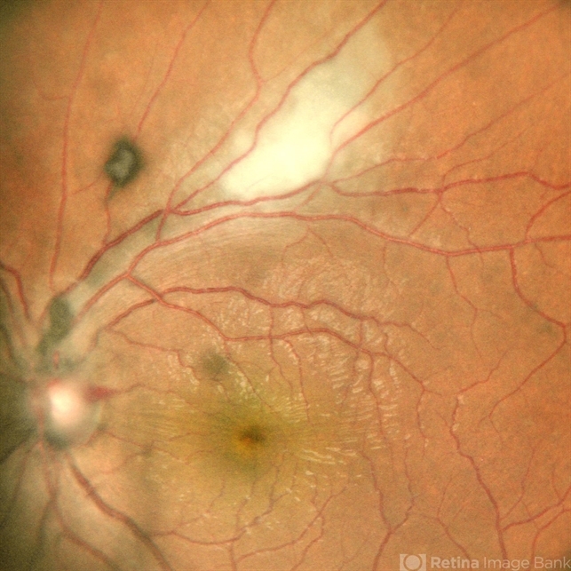
- toxoplasmosis retinitis
- Photographer
- DR. TEJASWITA VERMA
- Imaging device
-
Scanning laser ophthalmoscope
MIRANTE - Description
- Fundus photograph of the left eye of a 20 year old male with 6/12 vision showing a posterior pole fluffy yellowish white lesion along superotemporal arcade with full thickness involvement of retinal layers on OCT suggestive of toxoplasma retinitis .He was started on Tab. Bactrim-DS followed by oral steroids after 4 days .There was anterior segment involvement with few Keratic precipitates , IOP was 47mm Hg, therefore patient was misdiagnosed as viral trabeculitis elsewhere. IOP was managed medically.


---thumb.jpg/image-square;max$79,0.ImageHandler "Toxoplasmosis")
---thumb.jpg/image-square;max$79,0.ImageHandler "Acute Toxoplasmosis")
---thumb.jpg/image-square;max$79,0.ImageHandler "Acute Toxoplasmosis")


