-
 Central Retinal Artery Occlusion
Apr 10 2024 by Tejaswita Verma
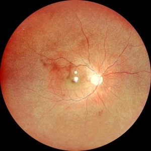
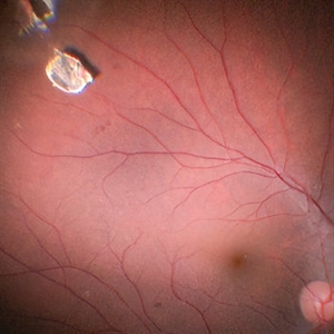
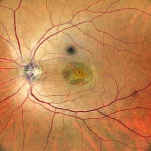
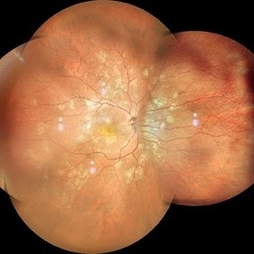
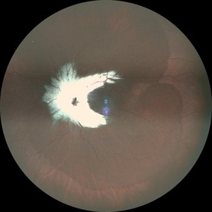
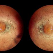
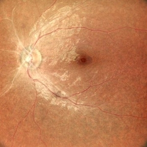
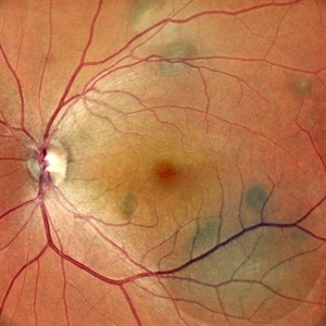
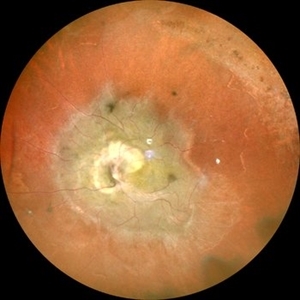
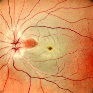
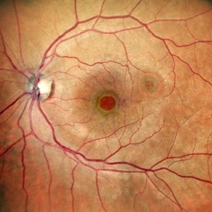
Left eye fundus photo of a 75 year old male with pale edematous retina with cherry red spot in a case of central retinal artery occlusion.
Photographer: DR. TEJASWITA VERMA
Imaging device: MIRANTE
Condition/keywords: central retinal artery occlusion (CRAO), cherry red spot
-
 Central Retinal Artery Occlusion OCT
Apr 10 2024 by Tejaswita Verma
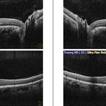
Left eye OCT of a 75 year old male with central retinal artery occlusion showing altered foveal contour with loss of differentiation of layers with thickening.
Photographer: DR. TEJASWITA VERMA
Imaging device: MIRANTE
Condition/keywords: central retinal artery occlusion, cherry red spot
-
 Central Retinal Artery Occlusion-Widefield
Apr 10 2024 by Tejaswita Verma
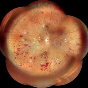
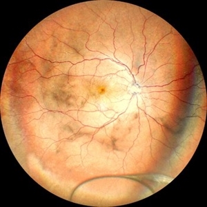
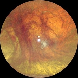
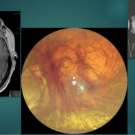
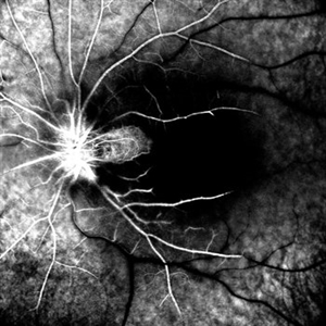
Left eye widefield color photo of 75 year old male with pale edematous retina with cherry red spot suggestive of central retinal artery occlusion.
Photographer: DR. TEJASWITA VERMA
Imaging device: MIRANTE
Condition/keywords: central retinal artery occlusion (CRAO), cherry red spot
-
 Tractional Retinal Detachment
Apr 20 2024 by Tejaswita Verma
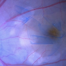
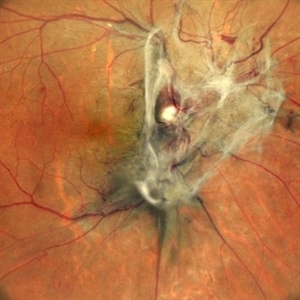
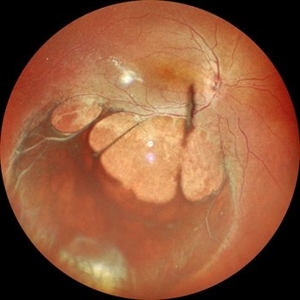
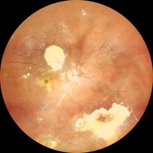
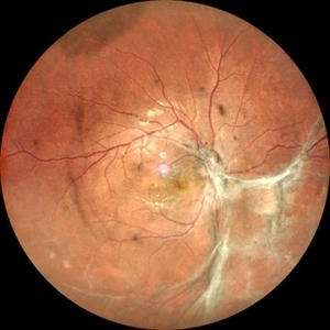
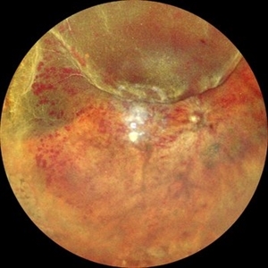
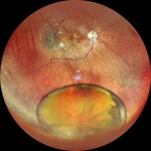
Fundus photograph of the right eye of a 62 year old female with tractional retinal detachment in a case of lasered proliferative diabetic retinopathy showing neovascularisation at disc and elsewhere
Photographer: DR. TEJASWITA VERMA
Condition/keywords: Neovascularisation at the Disc (NVD), Neovascularisation elsewhere (NVE), proliferative diabetic retinopathy (PDR)
-
 Proliferative Diabetic Retinopathy
Apr 20 2024 by Tejaswita Verma
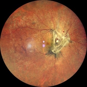
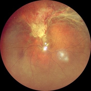
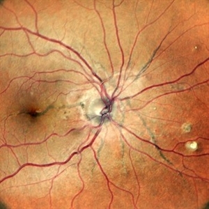
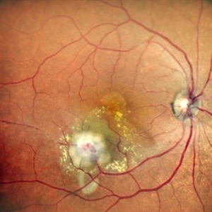
Fundus photograph of the left eye of a 62 year old female with lasered proliferative diabetic retinopathy showing neovascularisation elsewhere with few dot-blot haemorrhages
Photographer: DR. TEJASWITA VERMA
Condition/keywords: Neovascularisation elsewhere (NVE), pan-retinal photocoagulation (PRP), proliferative diabetic retinopathy (PDR)
-
 Proliferative Diabetic Retinopathy
Apr 20 2024 by Tejaswita Verma
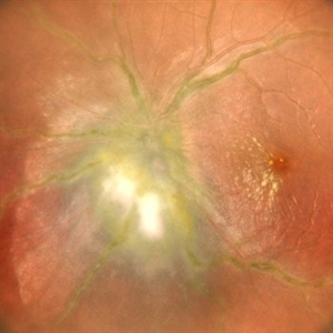
Widefield fundus photograph of the left eye of a 62 year old female with left eye lasered proliferative diabetic retinopathy showing neovascularisation elsewhere with few dot-blot hemorrhages.
Photographer: DR. TEJASWITA VERMA
Condition/keywords: Neovascularisation elsewhere (NVE), pan-retinal photocoagulation (PRP), proliferative diabetic retinopathy (PDR)
-
 Stargardt's Disease
Apr 20 2024 by Tejaswita Verma
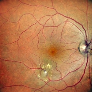
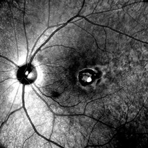
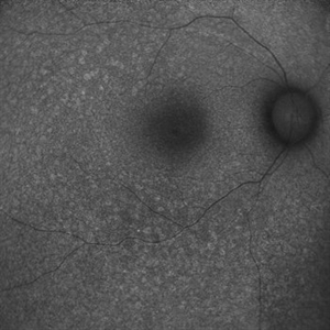
Fundus autofluorescence image of the right eye of a 39 year old male showing hypoautofluorescence in a case of Stargardt's disease.
Photographer: DR. TEJASWITA VERMA
Imaging device: MIRANTE
Condition/keywords: fundus autofluorescence (FAF), hereditary macular dystrophy, hypoautofluorescence, Stargardt disease
-
 Stargardt's Disease
Apr 20 2024 by Tejaswita Verma
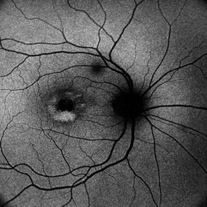
Fundus autofluorescence image of the left eye of a 39 year old male showing hypoautofluorescence in a case of Stargardt's disease.
Photographer: DR. TEJASWITA VERMA
Imaging device: MIRANTE
Condition/keywords: fundus autofluorescence (FAF), hereditary macular dystrophy, hypoautofluorescence, Stargardt disease
-
 Floating Ozurdex Implant
May 20 2024 by Tejaswita Verma
Fundus photograph of the left eye of a 73 year old female with ozurdex implant floating in the vitreous in a diabetic lasered patient.
Photographer: DR. TEJASWITA VERMA
Imaging device: MIRANTE
Condition/keywords: diabetic macular edema, laser photocoagulation, Ozurdex implant
-
 OCT in Subclinical RD in a Case of Retinitis Pigmentosa
May 22 2024 by Tejaswita Verma
OCT of a 6 year old male child showing subretinal fluid with macula off in case of subclinical retinal detachment in retinitis pigmentosa
Photographer: DR. TEJASWITA VERMA
Imaging device: MIRANTE
Condition/keywords: OCT, right eye, subclinical detachment
-
 OCT in Subclinical RD in a Case of Retinitis Pigmentosa
May 22 2024 by Tejaswita Verma
OCT of a 6 year old male child showing subretinal fluid with macula off in a case of subclinical RD in retinitis pigmentosa.
Photographer: DR. TEJASWITA VERMA
Imaging device: MIRANTE
Condition/keywords: left eye, OCT, retinitis pigmentosa, subclinical detachment
-
 Subclinical RD in Retinitis Pigmentosa
May 22 2024 by Tejaswita Verma
Fundus photograph of the left eye of a 6 year old male with subclinical retinal detachment with macula off in a case of retinitis pigmentosa with high myopia.
Photographer: DR. TEJASWITA VERMA
Imaging device: MIRANTE
Condition/keywords: high myopia, retinitis pigmentosa, subclinical detachment
-
 Subclinical RD in Retinitis Pigmentosa
May 22 2024 by Tejaswita Verma
Fundus photograph of the right eye of a 6 year old male with subclinical retinal detachment with macula off in case of retinitis pigmentosa with high myopia.
Photographer: DR. TEJASWITA VERMA
Imaging device: MIRANTE
Condition/keywords: high myopia, retinitis pigmentosa, subclinical detachment
-
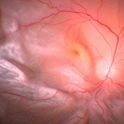
 ILM Peeling in Case of Optic Disc Pit Maculopathy
Jun 14 2024 by Tejaswita Verma
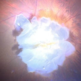
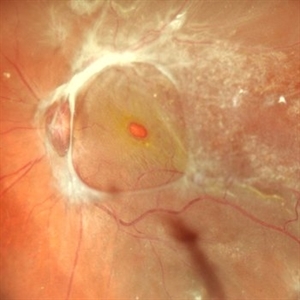
Intraoperative still of a 38 year old male post initiation of ILM peeling in a case of optic disc pit maculopathy.
Photographer: DR. TEJASWITA VERMA
Condition/keywords: intraoperative, optic pit
-
 Nucleus Drop
Jun 14 2024 by Tejaswita Verma
Intraoperative still of lens drop in a 63 year old female after an eventful cataract surgery.
Photographer: DR. TEJASWITA VERMA
Condition/keywords: intraoperative, lens matter, nucleus drop
-
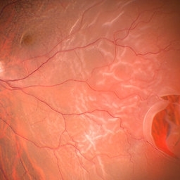
 Subretinal Silicon Oil With Inferior PVR
Jun 14 2024 by Tejaswita Verma
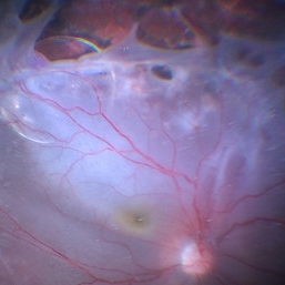
Intraoperative still of a 34 year old male with sub retinal silicon oil with proliferative vitreoretinopathy changes.
Photographer: DR. TEJASWITA VERMA
Condition/keywords: silicone oil
-
 IOFB Loosened Away From Imapct Site
Jun 14 2024 by Tejaswita Verma
Intraoperative still of a 21 year old male showing intraocular foreign body loosened away from the site of impact for easier removal.
Photographer: DR. TEJASWITA VERMA
Condition/keywords: intraocular foreign body, intraoperative
-
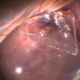 Glass Piece Inside Eye
Jun 14 2024 by Tejaswita Verma
Intraoperative still of a young male showing intraocular foreign body (glass piece) inside eye with retinal detachment.
Photographer: DR. TEJASWITA VERMA
Condition/keywords: intraocular foreign body
-
 Starfolds
Jun 14 2024 by Tejaswita Verma
Intraoperative still depicting starfolds in a case of retinal detachment with PVR changes.
Photographer: DR. TEJASWITA VERMA
Condition/keywords: starfolds
-
 Large Horseshoe Tear
Jun 14 2024 by Tejaswita Verma
Intraoperative still showing retinal detachment with a horse shoe tear.
Photographer: DR. TEJASWITA VERMA
-
 Atypical Tubercular Peripheral Occlusive Retinal Vasculitis
Jun 21 2024 by Tejaswita Verma
Fundus montage of the right eye of a 27 year old male with macula threatening occlusive vasculitis showing hemorrhages in inferior, temporal quadrant with vascular sheathing. The patient was Mantoux positive (20 mm induration) and IGRA (TB-GOLD)positive and started on oral steroids. The case was atypical due to no vitritis at presentation which is unusual of tuberculosis. Behcet's disease was ruled out as there was no panuveitis like picture.
Photographer: DR. TEJASWITA VERMA
Imaging device: MIRANTE
Condition/keywords: occlusive vasculitis, ocular tuberculosis
-
 FFA in Atypical Tubercular Peripheral Occlusive Retinal Vasculitis
Jun 21 2024 by Tejaswita Verma
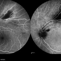
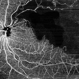
Right eye FFA montage of a 27 year male with peripheral occlusive tubercular vasculitis, showing CNP areas inferiorly and temporally, leakages and blocked fluorescence due to hemorrhages. The patient was advised intravitreal anti-VEGF injection and later sectoral laser once inflammation subsides.
Photographer: DR. TEJASWITA VERMA
Imaging device: MIRANTE
Condition/keywords: obliterative peripheral vasculitis, ocular tuberculosis
-
 Atypical Tubercular Occlusive Peripheral Retinal Vasculitis
Jun 21 2024 by Tejaswita Verma
Follow up right eye fundus photograph of a 27 year old male with vision 6/12 , diagnosed with systemic tuberculosis(mediastinal lymphadenopathy on chest CT) on oral steroids, and started on ATT .We can see a parafoveal sub-ILM hemorrhage with vascular sheathing and hemorrhages in inferior and temporal quadrants . The patient was advised anti-VEGF intravitreal injection, later sectoral laser after resolution of inflammation
Photographer: DR. TEJASWITA VERMA
Imaging device: MIRANTE
Condition/keywords: obliterative peripheral vasculitis, ocular tuberculosis
-
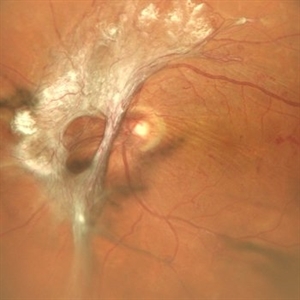
 Tractional Retinal Detachment
Jun 25 2024 by Tejaswita Verma
Fundus photograph of an elderly male showing diabetic tractional retinal detachment in a lasered eye.
Photographer: DR. TEJASWITA VERMA
Imaging device: MIRANTE
Condition/keywords: fibrovascular proliferation, proliferative diabetic retinopathy (PDR), tractional retinal detachment
-
 TRD
TRD
Jun 25 2024 by Tejaswita Verma
Right eye fundus photo of an elderly male having diabetic tractional retinal detachment in a case of lasered Proliferative diabetic retinopathy.
Photographer: DR. TEJASWITA VERMA
Imaging device: MIRANTE
Condition/keywords: laser photocoagulation, proliferative diabetic retinopathy (PDR), tractional retinal detachment
-
 RAM
RAM
Jun 25 2024 by Tejaswita Verma
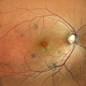
Right eye Fundus photo of a 73 year old female with 6/ 9 vision having retinal artery macroaneurysm.
Photographer: DR. TEJASWITA VERMA
Imaging device: MIRANTE
Condition/keywords: RETINAL ARTERY MACROANEURYSM
-
 Spontaneously Settled RRD
Jun 25 2024 by Tejaswita Verma
Fundus photograph of a 35 year old young male with 6/9 vision showing spontaneously settled inferior RD.
Photographer: DR. TEJASWITA VERMA
Imaging device: MIRANTE
Condition/keywords: Retinal detachment, spontaneous resolution
-
 Adult Vitelliform Macular Dystrophy
Jun 25 2024 by Tejaswita Verma
Left eye fundus photograph of an elderly 62 year old hypertensive female showing elevated lesion at macula s/o adult vitelliform macular dystrophy, misdiagnosed as long standing CSR elsewhere.
Photographer: DR. TEJASWITA VERMA
Imaging device: MIRANTE
Condition/keywords: Adult vitelliform macular dystrophy
-
 Adult Vitelliform Macular Dystrophy
Jun 25 2024 by Tejaswita Verma
Right eye Fundus photo of an elderly 62 year old female with 6/12 vision showing elevated lesion at macula with suggestive of adult vitelliform macular dystrophy, misdiagnosed as long standing CSR elsewhere.
Photographer: DR. TEJASWITA VERMA
Imaging device: MIRANTE
Condition/keywords: Adult-onset vitelliform dystrophy
-
 OCT in Adult Vitelliform Dystrophy
Jun 25 2024 by Tejaswita Verma
OCT image of a 62 year old female with 6/12 vision in both eyes showing sub retinal fluid with RPE granularity s/o Adult vitelliform macular dystrophy.
Photographer: DR. TEJASWITA VERMA
Imaging device: MIRANTE
Condition/keywords: adult vitelliform dystrophy, optical coherence tomography (OCT)
-
 B-FAF in Stargardt's Disease
Jul 4 2024 by Tejaswita Verma
Blue fundus autofluorescence showing hypoautofluorescence picture of a 28 year old male with 6/60 vision in BE in a case of Stargardt's disease.
Photographer: DR. TEJASWITA VERMA
Imaging device: MIRANTE
Condition/keywords: fundus autofluorescence (FAF), hereditary macular dystrophy, Stargardt disease
-
 Retinal Artery Macroaneurysm
Jul 13 2024 by Tejaswita Verma
A 53 year old female presented with blurred vision in RE since a month ,with borderline DM and HTN not on medications .H/o highest BP recording was 160/90 mm Hg.Vision 6/60 .FFA revealed leakages. She was advised RE focal laser with intravitreal anti-VEGF injections
Photographer: DR. TEJASWITA VERMA
Imaging device: MIRANTE
Condition/keywords: RETINAL ARTERY MACROANEURYSM
-
 Multifocal Choroiditis
Jul 13 2024 by Tejaswita Verma
RE fundus montage of a 34 y/o male showing old and active hypopigmented lesions with macular involvement .He presented with DOV since a month,treated with oral steroids for 15 days elsewhere,with BCVA of CF2mt and positive Mantoux test.
Photographer: DR. TEJASWITA VERMA
Imaging device: MIRANTE
Condition/keywords: multifocal choroiditis
-
 Benign Familial Fleck Retina
Jul 13 2024 by Tejaswita Verma
Fundus photograph of the LE of a 46 y/o female with bilateral diffuse yellow-white fleck like lesions with 6/6 vision bilaterally.
Photographer: DR. TEJASWITA VERMA
Imaging device: MIRANTE
Condition/keywords: Benign familial fleck retina, flecks
-
 Benign Familial Fleck Retina
Jul 13 2024 by Tejaswita Verma
Fundus photograph of the RE of a 46 y/o female with bilateral diffuse yellow-white fleck like lesions with 6/6 vision bilaterally
Photographer: DR. TEJASWITA VERMA
Imaging device: MIRANTE
Condition/keywords: Benign familial fleck retina
-
 Benign Familial Fleck Retina
Jul 13 2024 by Tejaswita Verma
Fundus photograph of the LE of a 46 y/o female with bilateral diffuse yellow-white fleck like lesions with 6/6 vision bilaterally
Photographer: DR. TEJASWITA VERMA
Imaging device: MIRANTE
Condition/keywords: Benign familial fleck retina
-
 Benign Familial Fleck Retina
Jul 13 2024 by Tejaswita Verma
Fundus photograph of the RE of a 46 y/o female with bilateral diffuse yellow-white fleck like lesions with 6/6 vision bilaterally.
Photographer: DR. TEJASWITA VERMA
Imaging device: MIRANTE
Condition/keywords: Benign familial fleck retina, flecks
-
 OCT Line Scan in Benign Familial Fleck Retina
Jul 13 2024 by Tejaswita Verma
OCT line scan in flecked retina with foveal involvement, showing slightly thickened RPE with multiple PEDs and intact IS-OS junction with 6/6 vision bilaterally.
Photographer: DR. TEJASWITA VERMA
Imaging device: MIRANTE
Condition/keywords: Benign familial fleck retina
-
 Foveoschisis
Jul 31 2024 by Tejaswita Verma
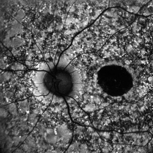
Fundus autofluorescence image of a 17 year old boy showing spokewheel pattern bilaterally in case of foveoschisis
Photographer: DR. TEJASWITA VERMA
Imaging device: MIRANTE
Condition/keywords: foveoschisis
-
 Juvenile Foveoschisis
Jul 31 2024 by Tejaswita Verma
Central fundus image of a teenage boy with 6/12 vision showing spokewheel pattern in case of juvenile X-linked bilateral foveoschisis.
Photographer: DR. TEJASWITA VERMA
Imaging device: MIRANTE
Condition/keywords: foveoschisis, juvenile retinoschisis
-
 Juvenile X-linked Retinoschisis
Jul 31 2024 by Tejaswita Verma
Widefield fundus image of a 17 year old boy with BCVA 6/12 showing juvenile X-linked peripheral retinoschisis and foveoschisis bilaterally.
Photographer: DR. TEJASWITA VERMA
Imaging device: MIRANTE
Condition/keywords: juvenile retinoschisis
-
 RD with PVR in CMV Retinitis in an HIV Positive Patient
Jul 31 2024 by Tejaswita Verma
Fundus photograph of a 48 year old male with CF 1.5 mt vision having history of CMV retinitis, on HAART with CD4 count 81, showing retinal detachment with proliferative vitreoretinopathy changes. He was advised pars plana vitrectomy with silicon oil infusion.
Photographer: DR. TEJASWITA VERMA
Imaging device: MIRANTE
Condition/keywords: CMV retinitis with retinal detachment, HIV
-
 Post-Fever Retinitis
Jul 31 2024 by Tejaswita Verma
Fundus image of a young 19 year old female with a week old history of fever and CF vision showing disc edema and macular star. She was advised Weil Felix test but lost to follow up.
Photographer: DR. TEJASWITA VERMA
Imaging device: MIRANTE
Condition/keywords: POST FEVER RETINITIS
-
 Myelinated Nerve Fibres With Combined Hamartoma of Retina and RPE
Jul 31 2024 by Tejaswita Verma
Fundus image of a 20 year old female who presented with metamorphopsia ,slightly blurred vision. BCVA was 6/9, epiretinal membrane present on central fundus examination with myelinated nerve fibres.
Photographer: DR. TEJASWITA VERMA
Imaging device: MIRANTE
Condition/keywords: combined hamartoma of retina and RPE, myelinated nerve fibers
-
 Signet Ring in the Eye
Sep 28 2024 by Tejaswita Verma
Infrared fundus image of the LE of a 32 year-old male showing macular coloboma.
Photographer: DR. TEJASWITA VERMA
Imaging device: MIRANTE
Condition/keywords: macular coloboma
-
 Combined Retinal Detachment With Macular Hole
Sep 28 2024 by Tejaswita Verma
Fundus image of the LE of a 67 year old diabetic, hypertensive female with CF 3metres vision showing combined RD with FTMH, in a pseudophakic eye. She was lost to follow up status post 2 anti VEGF injections received 8 months back due to typhoid fever.
Photographer: DR. TEJASWITA VERMA
Imaging device: MIRANTE
Condition/keywords: full thickness macular hole, proliferative diabetic retinopathy (PDR), tractional retinal detachment
-
 Adult Onset Vitelliform Macular Dystrophy
Sep 29 2024 by Tejaswita Verma
Autofluorescence image of the RE of a 62 year-old hypertensive female with 6/12 vision showing hyperautofluorescence . The patient was asked to review every few months to check for development of secondary CNVM
Photographer: DR. TEJASWITA VERMA
Imaging device: MIRANTE
Condition/keywords: Adult vitelliform macular dystrophy
-
 Retinitis Pigmentosa With Papilledema
Sep 29 2024 by Tejaswita Verma
Fundus pictures of RE and LE of a 30 year old male with a 20 day history of diminution of vision in LE more than RE. Vision was 6/9P and 6/24P in RE and LE respectively. History of RP in father. Alleged history of trauma to left side of forehead 4 months back with subgaleal haematoma over left side of frontal region on CT done 4 months back, No significant intracranial abnormality. He was started on oral steroids with tapering and F/U after 2 weeks.
Photographer: DR. TEJASWITA VERMA
Imaging device: MIRANTE
Condition/keywords: papilledema, retinitis pigmentosa
-
 Coats Disease
Sep 29 2024 by Tejaswita Verma
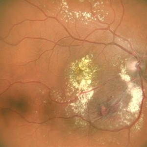
Fundus photo of the RE of a 14 y/o female ,nil premorbid presented with reduced vision in the RE ,diagnosed incidentally on ophthalmological examination elsewhere .Vision was finger counting 3 meters in the RE . Fundus picture reveals macular scar , subretinal and intraretinal exudation ,with scattered hemorrhages esp. in STQ, sclerosed vessels in superior, superonasal quadrant ,nasal, inferonasal quadrant, CR scars inferiorly, Telengiectatic vessels S/O Coat's disease. She was advised RE anti VEGF x1 + laser PRP + PST kenacort under GA with guarded prognosis.
Photographer: DR. TEJASWITA VERMA
Imaging device: MIRANTE
Condition/keywords: Coats' disease
-
 Angioid Streaks
Sep 29 2024 by Tejaswita Verma
Fundus photograph of a 35 year-old female with 6/6 vision in RE , unremarkable anterior segment and family history of angioid streaks and pseudoxanthoma elasticum in sister. Fundus examination revealed angioid streaks radiating from disc , sparing the fovea .Her Sister had received multiple anti VEGF injections for angioid streaks with CNVM.
Photographer: DR. TEJASWITA VERMA
Imaging device: MIRANTE
Condition/keywords: angioid streaks
-
 Toxoplasma Retinitis
Nov 4 2024 by Tejaswita Verma
Fundus photograph of the left eye of a 20 year old male with 6/12 vision showing a posterior pole fluffy yellowish white lesion along superotemporal arcade with full thickness involvement of retinal layers on OCT suggestive of toxoplasma retinitis .He was started on Tab. Bactrim-DS followed by oral steroids after 4 days .There was anterior segment involvement with few Keratic precipitates , IOP was 47mm Hg, therefore patient was misdiagnosed as viral trabeculitis elsewhere. IOP was managed medically.
Photographer: DR. TEJASWITA VERMA
Imaging device: MIRANTE
Condition/keywords: toxoplasmosis retinitis
-
 Proliferative Retinopathy
Nov 4 2024 by Tejaswita Verma
Fundus photograph of a middle aged male with diabetes showing large FVP following NVD.
Photographer: DR. TEJASWITA VERMA
Imaging device: MIRANTE
Condition/keywords: FVPs, neovascularization of the disc (NVD), proliferative diabetic retinopathy (PDR)
-
 Atypical Retintis Pigmentosa With Old CRAO
Nov 15 2024 by Tejaswita Verma
Fundus image of a 20 year old female who presented with bilateral visual loss since 2 months following history of typhoid fever, UTI, showing pale disc, sclerosed vessels,altered foveal reflex and granular fundus. Vision was light perception in both eyes. CBC, MRI Brain +Orbit , carotid doppler tests were WNL. Brucella IgG,IgM negative. ERG and VEP were abnormal.
Photographer: DR. TEJASWITA VERMA
Imaging device: MIRANTE
Condition/keywords: ATYPICAL RETINITIS PIGMENTOSA, CRAO
-
 Dislocated Intraocular Lens
Nov 15 2024 by Tejaswita Verma
Fundus image of a spontaneously posteriorly dislocated IOL 10 years following surgery. Other eye had a subluxated opacified IOL.
Photographer: DR. TEJASWITA VERMA
Imaging device: MIRANTE
Condition/keywords: dislocated intraocular lens (IOL)
-
 Venolymphatic Mass with Retinal Folds
Nov 25 2024 by Tejaswita Verma
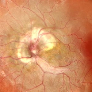
Fundus picture of a 26 year old male who presented with right eye abaxial proptosis, MRI confirmed venolymphatic mass inferomedial in location located near the optic disc with disc edema , nasal elevation ,retinal folds. Vision was 6/18 . He was planned for intralesional bleomycin injection.
Photographer: DR. TEJASWITA VERMA
Imaging device: MIRANTE
Condition/keywords: disc edema, intraorbital mass, proptosis, retinal folds
-
 Venolymphatic Mass With Disc Edema
Dec 5 2024 by Tejaswita Verma
Fundus picture of a 26 year old male who presented with right eye abaxial proptosis, MRI confirmed venolymphatic mass inferomedial in location located near the optic disc with disc edema , nasal elevation ,retinal folds. Vision was 6/18 . He was planned for intralesional bleomycin injection.
Photographer: DR. TEJASWITA VERMA
Imaging device: MIRANTE
Condition/keywords: disc edema, intraorbital mass, proptosis
-
 Atypical RP with Typhoid Retinitis Sequelae with Old CRAO
Dec 5 2024 by Tejaswita Verma
FAF of a 20 year old female who presented with 2 months history of sudden painless vision loss, bilaterally light perception vision, s/o presumed atypical RP, bilateral old CRAO with typhoid retinitis sequelae.
Photographer: DR. TEJASWITA VERMA
Imaging device: MIRANTE
Condition/keywords: CRAO, retinitis pigmentosa, typhoid fever
-
 Multiple PEDs
Dec 5 2024 by Tejaswita Verma
Fundus photo of a 40 year old female with Multiple PEDs ,vision was 6/9 . Other eye had CSR.
Photographer: DR. TEJASWITA VERMA
Imaging device: MIRANTE
Condition/keywords: pigment epithelial detachment (PED)
-
 CSR with multiple PEDs
Dec 5 2024 by Tejaswita Verma
Fundus picture of the right eye of a 40 year old female with 6/12 vision showing exudative subretinal fluid and multiple PEDs in a case of CSR.
Photographer: DR. TEJASWITA VERMA
Imaging device: MIRANTE
Condition/keywords: central serous retinopathy (CSR), pigment epithelial detachment (PED)
-
 Toxoplasmosis
Dec 5 2024 by Tejaswita Verma
26 year old male with 6/18 vision , anterior chamber reaction, vitritis and retinitis lesion along the superotemporal arcade with full thickness involvement on OCT . FFA showing hypofluorescence with surrounding hyperfluorescence characterstic of toxoplasma retinitis . ICGA shows hypocyanescence.
Photographer: DR. TEJASWITA VERMA
Imaging device: MIRANTE
Condition/keywords: Fundus Fluorescein Angiography, indocyanine green (ICG) angiography, toxoplasmosis
-
 Extramacular TRD in Idiopathic Occlusive Vasculitis
Dec 5 2024 by Tejaswita Verma
Fundus photo showing extramacular TRD in a 16 year old boy with idiopathic occlusive vasculitis secondary to presumed IOTB. History of taking ATT for 6 months , Mantoux positive previously. Vision was 6/6P,other eye had funnel RD .
Photographer: DR. TEJASWITA VERMA
Imaging device: MIRANTE
Condition/keywords: tractional retinal detachment, vasculitis
-
 PDR with NVD
Dec 5 2024 by Tejaswita Verma
Fundus image of a middle aged male with NVD, multiple dot blot and flame shaped hemorrhages, cotton wool spots, hard exudates at the posterior pole in a case of PDR . Vision was 6/9.
Photographer: DR. TEJASWITA VERMA
Imaging device: MIRANTE
Condition/keywords: NEOVASCULARISATION OF DISC, proliferative diabetic retinopathy (PDR)
-
 Unilateral Coloboma Involving Disc and Macula
Dec 27 2024 by Tejaswita Verma
Fundus image of a 15 years old male presenting with unilaterally diminished vision since childhood in RE with CF3mt vision and inferior iris coloboma and retinochoroidal coloboma with nystagmus and cataract.
Photographer: DR. TEJASWITA VERMA
Imaging device: MIRANTE
Condition/keywords: chorioretinal coloboma, iridofundal coloboma
-
 Polyploidal Choroidal Vasculopathy
Dec 27 2024 by Tejaswita Verma
Fundus image of a 74 year old woman with CF1mt vision in right eye showing large PED in a case of PCV. There was associated full thickness macular hole in the same eye.
Photographer: DR. TEJASWITA VERMA
Imaging device: MIRANTE
Condition/keywords: PED, polypoidal choroidal vasculopathy (PCV)
-
 Post Dengue Fever Retinitis
Dec 27 2024 by Tejaswita Verma
A 35 year old female presented with bilaterally decreased vision since a month post dengue fever. Vision was finger counting 6 mt. in the RE and 6/36 in the left eye .Fundus examination revealed severe vitritis in the RE with retinitis lesions and hemorrhages and macular star ,Fundus examination of the left eye revealed moderate vitritis with retinitis lesions with hemorrhages and macular star. She was started on tab doxycycline 100 mg BD for 2 weeks and oral steroids
Photographer: DR. TEJASWITA VERMA
Imaging device: MIRANTE
Condition/keywords: Dengue Fever, macular star, POST FEVER RETINITIS
-
 Post Dengue Fever Retinitis
Dec 27 2024 by Tejaswita Verma
A 35 year old female presented with bilaterally decreased vision since a month post dengue fever. Vision was finger counting 6 mt. in the RE and 6/36 in the left eye .Fundus examination revealed severe vitritis in the RE with retinitis lesions and hemorrhages and macular star ,Fundus examination of the left eye revealed moderate vitritis with retinitis lesions with hemorrhages and macular star. She was started on tab doxycycline 100 mg BD for 2 weeks and oral steroids. She was also advised RE intravitreal implant Ozurdex with explained prognosis.
Photographer: DR. TEJASWITA VERMA
Imaging device: MIRANTE
Condition/keywords: bilateral dengue retinitis, Dengue Fever, POST FEVER RETINITIS
-
 Posterior Scleritis
Jan 4 2025 by Tejaswita Verma
Left eye fundus photo of a 49 year old female presenting with 5 days history of blurred vision, Vision was finger counting 2 mts. and nodular posterior scleritis with T sign on USG was present. OCT revealed altered foveal contour with septations and SRF pockets and bacillary layer detachment.
Photographer: DR. TEJASWITA VERMA
Imaging device: MIRANTE
Condition/keywords: posterior scleritis
-
 Combined Hamartoma of the Retina and RPE
Jan 23 2025 by Tejaswita Verma
A 10 year old boy presented with 6/60 vision and LE exotropia with the fundus lesion suggesting a chronic etiology and ILM folds.
Photographer: DR. TEJASWITA VERMA
Imaging device: MIRANTE
Condition/keywords: combined hamartoma of retina and RPE
-
 Choroidal Melanoma Masquerading as Subretinal Hemorrhage With Breakthrough VH
Jan 23 2025 by Tejaswita Verma
A 65 year old diabetic male presented with large nasal retinal mass giving the appearance of organized dehaemoglobinized subretinal hemorrhage with breakthrough vitreous hemorrhage , with 6/6P vision. Enucleation specimen showed histopathology confirmed choroidal melanoma.
Photographer: DR. TEJASWITA VERMA
Imaging device: MIRANTE
-
 Leber's Miliary Aneurysm
Jan 23 2025 by Tejaswita Verma
A 41 year old male presented with 6/9 vision in the RE and fundus picture revealed miliary aneurysm with exudates and hemorrhages surrounded by old focal and sectoral laser marks. OCT revealed altered foveal contour with cystic spaces and IRHRM. He was advised RE injection antiVEGF with focal laser.
Photographer: DR. TEJASWITA VERMA
Imaging device: MIRANTE
Condition/keywords: Leber's miliary aneurysm
-
 Coat's Disease with Exudative RD
Feb 12 2025 by Tejaswita Verma
Fundus photo of a 7 year old boy with vision Counting fingers close to face in the right eye and intermittent outward deviation of the right eye observed by parents. Fundus examination shows subretinal exudates, telengiectatic vessels in superotemporal quadrant, intraretinal hemorrhages, macular scar, NVD.
Photographer: DR. TEJASWITA VERMA
Imaging device: MIRANTE
Condition/keywords: Coats' disease, exudative retinal detachment
-
 Choroidal Melanoma Masquerading as PEHCR
Mar 3 2025 by Tejaswita Verma
A 65 year old diabetic male presented with large nasal retinal mass giving the appearance of organized dehaemoglobinized subretinal hemorrhage with breakthrough vitreous hemorrhage , with 6/6P vision. Enucleation specimen showed histopathology confirmed choroidal melanoma.
Photographer: DR. TEJASWITA VERMA
Imaging device: MIRANTE
Condition/keywords: vitreous hemorrhage
-
 Intravitreal Ozurdex Implant
Apr 3 2025 by Tejaswita Verma
Fundus photo of a middle-aged diabetic male showing Ozurdex implant in situ with laser marks.
Photographer: Tejaswita Verma
Imaging device: MIRANTE
Condition/keywords: dexamethasone implant, ozurdex
-
 Macular Schisis with RD
Apr 4 2025 by Tejaswita Verma
Fundus photo of a 29 year-old male with RRD, breaks in periphery, macular schisis . Vision CF2.5 mts
Photographer: DR. TEJASWITA VERMA
Imaging device: MIRANTE
Condition/keywords: macular schisis, retinal detachment
-
 Large Horseshoe Tear
Apr 4 2025 by Tejaswita Verma
Fundus photo of a 29 year old myopic male with RE 6/12P vision with large ragged tear on the posterior edge of lattice degeneration
Photographer: DR. TEJASWITA VERMA
Imaging device: MIRANTE
Condition/keywords: horseshoe tear
-
 Hereditary Macular Dystrophy with CNVM
Apr 4 2025 by Tejaswita Verma
Fundus photo of a 46 y/o male with 6/36 vision, Stargardt's disease with CNVM
Photographer: DR. TEJASWITA VERMA
Imaging device: MIRANTE
Condition/keywords: CNVM, hereditary macular dystrophy, HMD
-
 Neovascularisation Disc
Apr 4 2025 by Tejaswita Verma
Fundus photo of a 38 year-old Type 1 diabetic male with 6/18P vision in RE, presenting with TRD, NVD.
Photographer: DR. TEJASWITA VERMA
Imaging device: MIRANTE
Condition/keywords: neovascularization of the disc (NVD), TRD
-
 CRVO with RD
Apr 4 2025 by Tejaswita Verma
Fundus photo of a 58 year-old hypertensive male who presented with RE 6/60 vision with CRVO, rhegmatogenous RD.H/O DOV in RE since 3 days, H/O receiving anti VEGF X 2 injections 2 months ago
Photographer: DR. TEJASWITA VERMA
Imaging device: MIRANTE
Condition/keywords: central retinal vein occlusion (CRVO), Retinal Detachment
-
 CRAO Sparing Cilioretinal Artery
May 1 2025 by Tejaswita Verma
Fundus photo of a middle aged male with 6/60 vision in left eye showing CRAO partially sparing cilioretinal artery.
Photographer: Dr. Tejaswita Verma
Imaging device: MIRANTE
Condition/keywords: cilioretinal sparing, CRAO
-
 Cilioretinal Artery Sparing CRAO
May 1 2025 by Tejaswita Verma
Fundus photo of a middle aged male with CRAO partially sparing cilioretinal artery and papillomacular bundle. Vision 6/60.
Photographer: Dr. Tejaswita Verma
Imaging device: MIRANTE
Condition/keywords: CRAO with cilioretinal sparing
-
 Dark Territories
Jun 27 2025 by Tejaswita Verma
OCT-Angio image of the LE of a 76 year old hypertensive male with history of old superotemporal branch retinal vein occlusion status post 3 anti VEGF injections received in 2012.Vision was 6/9 in left eye. OCTA shows a large CNP area .
Photographer: Dr. Tejaswita Verma
Imaging device: MIRANTE
Condition/keywords: branch retinal vein occlusion (BRVO), CNP areas, OCTA, ST BRVO
-
 Double Trouble
Jun 28 2025 by Tejaswita Verma
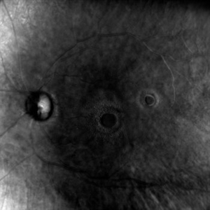
Retro image of aa double macular hole in a 60 yr old diabetic female status post PPV + gas 4 months ago. Vision was 6/60 in LE.
Photographer: Dr. Tejaswita Verma
Imaging device: MIRANTE
Condition/keywords: macular hole, retro mode
-
 Double Trouble
Jun 28 2025 by Tejaswita Verma
Fundus image of a 60 year old diabetic female with double macular holes with 6/60 vision status post LE PPV+gas 4 months ago. Other eye also had an unoperated large macular hole. Known case of glaucoma
Photographer: Dr. Tejaswita Verma
Imaging device: MIRANTE
Condition/keywords: macular hole
-
 The Horseshoe Of Havoc
Jun 28 2025 by Tejaswita Verma
Fundus image of a 50 year old male with a very large horseshoe tear causing RRD with macula off, hydration folds.
Photographer: DR. TEJASWITA VERMA
Imaging device: MIRANTE
Condition/keywords: horseshoe tear
-
 The Horseshoe Of Havoc
Jun 28 2025 by Tejaswita Verma
Fundus image of a 50 year old male with a very large horseshoe tear causing RRD with macula off, hydration folds.
Photographer: Dr. Tejaswita Verma
Imaging device: MIRANTE
Condition/keywords: horseshoe tear, large break
-
 Multiple HST Causing Subtotal RD
Jul 24 2025 by Tejaswita Verma
Fundus image of a 64 year old male with HM vision with pseudophakic bullous RD.
Photographer: Dr. Tejaswita Verma
Imaging device: MIRANTE
Condition/keywords: horseshoe tear, RD
-
 Retinal Artery Macroaneurysm With Macular Edema
Sep 12 2025 by Tejaswita Verma
Fundus photo of a 73 year old hypertensive female with 6/18 vision, presenting with RAM ,with surrounding hard exudates and macular edema. She was advised focal laser, anti VEGF injection.
Photographer: DR. TEJASWITA VERMA
Imaging device: MIRANTE
Condition/keywords: RAM, retinal arterial macroaneurysm
-
 Dislocated Nucleus
Sep 12 2025 by Tejaswita Verma
Fundus photo of a middle aged male with 6/36 vision, spontaneously dislocated nucleus posteriorly with focal retinal detachment. Right eye Pars plana Vitectomy + nucleus removal + intravitreal C3F8 (12%) gas was performed for this patient.
Photographer: DR. TEJASWITA VERMA
Imaging device: MIRANTE
Condition/keywords: dislocated lens, retinal detachment
-
 Lasered Retinal Artery Macroaneurysm
Sep 22 2025 by Tejaswita Verma
Fundus image of a 73 year old hypertensive female status post focal laser for exudative RAM. There was associated macular edema on OCT. Vision was 6/18.Patient was also planned for intravitreal anti VEGF injection on the same day.
Photographer: DR. TEJASWITA VERMA
Imaging device: MIRANTE
Condition/keywords: focal laser, RAM, retinal artery macroaneurysm
-
 A Dramatic Curtain Call in the Retina
Oct 13 2025 by Tejaswita Verma
Fundus image of a 15 year old boy with 3 day history of sudden DOV, IOP 6 mm Hg, HM vision showing nearly 270 degree GRT
Photographer: DR. TEJASWITA VERMA
Imaging device: MIRANTE
Condition/keywords: giant retinal tear
-
 Retinoblastoma
Oct 13 2025 by Tejaswita Verma
Fundus image of a 9 month old baby girl with normal bith history,who presented with leucocoria for 3 months bilaterally,USG BE was s/o calcification with endophytic RB.The image was taken in flying baby position under EUA
Photographer: Dr. Tejaswita Verma
Imaging device: MIRANTE
Condition/keywords: retinoblastoma
-
 Choroidal Mass
Jan 7 2026 by Tejaswita Verma
Suspected Choroidal mass in a 70 year old female diagnosed with PCV with subretinal hemorrhage elsewhere status post anti VEGF x 3 injections and gas displacement.
Photographer: Dr. Tejaswita Verma
Imaging device: MIRANTE
Condition/keywords: choroidal mass, subretinal hemorrhage
-
 Parafoveal Hyperautofluorescence in CSR
Jan 7 2026 by Tejaswita Verma
B-FAF of a 40 year old male with B-FAF showing hyperAF and smokestack leak on FFA .Vision 6/12
Photographer: DR. TEJASWITA VERMA
Imaging device: MIRANTE
Condition/keywords: Central Serous Chorioretinopathy (CSR), fundus autofluorescence
A project from the American Society of Retina Specialists