 Initializing download.
-
 By AVIK DEY SARKAR, MS, FVRS, FAICO(VR)
Kamala Sundaram Eye Center, Chembur, Mumbai, India
Co-author(s): Dr. Naresh Babu Kannan, MS, FVRS, MBA, FASRS, Chief, Dept. of Vitreoretinal Services, Aravind Eye Hospital, Madura, India - Uploaded on Oct 31, 2024.
- Last modified by Joshua Friedman on Oct 31, 2024.
- Rating
- Appears in
- 31-Oct-2024
- Condition/keywords
- retina, vascular anomaly, Diabetic Retinopathy, background diabetic retinopathy (BDR)
- Photographer
- Dr. Avik Dey Sarkar, MBBS, MS, FVRS, FAICO, Consultant, Department of Vitreoretinal Services, Aravind Eye Hospital, Madurai, India
- Imaging device
-
Fundus camera
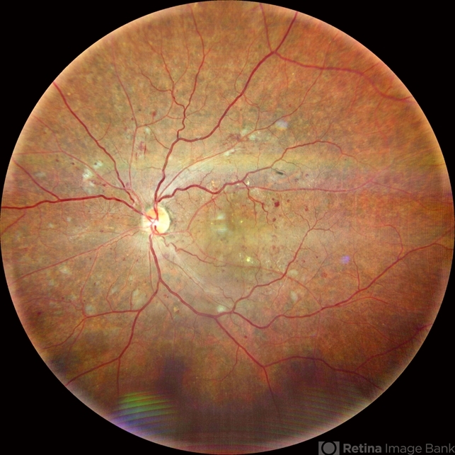
Wide angled Fundus imaging with Clarus 300 - Description
- A 43-year-old hypertensive patient, diagnosed with Non-Ischemic Central retinal vein Occlusion in OS, presented with a striking anatomical variation in retinal vasculature. The inferior first-order retinal arteriole after initiating from the optic disc bifurcates, before reaching the fovea, and the superior branch after crossing the midline forms the superior arcade afterwards and produces dichotomous branching as usual. This defies basic anatomical considerations for retinal vasculature as they never cross the midline, also known as the watershed line for retinal vessels.1,2 References: 1. May CA, Rutkowski P. The Horizontal Raphe of the Human Retina and its Watershed Zones. Vision. 2019; 3(4):60. 2. May CA, Rutkowski P. Hypothesis: watershed zones in the human eye are a key for understanding glaucomatous retinal damage. Med Hypotheses. 2017;109:1-5.


---thumb.jpg/image-square;max$79,0.ImageHandler "Retinoblastoma To Chemothermotherapy")
---thumb.jpg/image-square;max$79,0.ImageHandler "Retinoblastoma To Chemothermotherapy")


